A brief insight on ACE receptors role in COVID-19

A brief insight on ACE receptors role in COVID-19

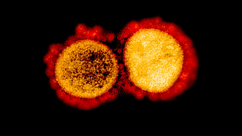
With the novel coronavirus, COVID-19 is spreading across the world and the fact that no drug or treatment has been found against it is creating fear among people. Coronavirus is a large family of enveloped, single-stranded RNA that infects mainly mammals including humans. In humans, coronavirus causes mild to severe respiratory illness. These viruses exhibit strong virulence and are highly contagious. While a person infected, produces mild symptoms, certain individuals respond severely, sometimes leading to death. 

The SARS pandemic, back in 2002 is known to belong to the same family of viruses as COVID-19. According to WHO, SARS rapidly spread through 29 countries, with 8096 confirmed cases, but the current pandemic has surpassed all numbers by infecting over millions of people across the world. Today, it is due to SARS that has resulted in a coordinated effort to develop treatments targeting the virus or host cell components responsible for viral replication.

The deadly virus, COVID-19 enters the human body and binds to the target cells through angiotensin-converting enzyme 2 (ACE2) which is mainly expressed in endothelial cells and Leydig cells. ACE-2, a transmembrane metallo carboxypeptidase, is an enzyme which for years has been important for the treatment of hypertension. With further Polymerase Chain Reaction (PCR) analysis it was determined that the ACE-2 receptor is also present in the lung and gastrointestinal tract, tissues harboring COVID-19. 

Inhibiting ACE2 receptor blocks COVID-19 entry:

ACE-2 belonging to the family of ACE receptors, plays a key role in the Renin-Angiotensin System (RAS) and in the treatment of hypertension. ACE2 is known to degrade angiotensin II and thereby, negatively regulating RAS. Recently experts have revealed that the COVID-19 virus uses ACE-2 receptors as their entry in HeLa cells. Additionally, it was found that by using anti- ACE-2 antibodies in other mammals like monkeys, it was seen that there was an entry blockage of pseudotypes expressing the COVID-19 virus. 

For the virus to enter the host cell, its spike glycoprotein (S) needs to be cleaved at 2 sites, termed S protein primming so the viral and host cells membrane can fuse. A serine protease TMPRSS2 is essential for cleaving the S protein. It was found by treating Calu-3 human lung cell line with a serine inhibitor- camostat mesylate the entry of the COVID-19 virus was partially blocked. Angiotensin-converting enzyme (ACE) inhibitors is also found to play a role in preventing the formation of angiotensin II. ACE multifaceted functions include treating heart failure, controlling high blood pressure, and preventing kidney failure in diabetic patients. 

Existing concerns about ACE inhibitors:

Though ACE inhibitors might seem like a promising solution for COVID-19, there are certain concerns regarding the increased susceptibility to COVID-19. These are considered based on the fact that ACE inhibitors are also used in treating millions of people with hypertension, heart, and kidney disease. When administered with inhibitors, diabetes and hypertension patients were observed to have an increase in ACE2, which in turn would facilitate infection with COVID-19. It is hence hypothesized that treating diabetic and hypertension patients with ACE inhibitors might make them more susceptible to COVID-19. 

Furthermore, several studies have reported that long term usage of ACE inhibitors can modify the adaptive immune response, which is a key and much-needed defense against any infection. These particular effects must be taken into noticed and investigated further in context to COVID-19. 

Road to COVID-19 therapies:

These findings could greatly impact the efforts being taken in developing treatments for the current pandemic. For instance, TMPRSS2 inhibitors can be potentially used to prevent virus entry into the host cells. Though there are certain drawbacks in using ACE inhibitors, there is a lack of scientific evidence and clinical data to support the discontinuing of ACE inhibitors in COVID-19 patients with existing hypertension and diabetes. The proof that reduction in mortality due to ACE inhibitors and the beneficial effects outweigh the theoretical risks. 

Our interpretation of this hypothesis should not lead to changing drugs for patients with diabetes or hypertension, without consulting an expert physician. Though it’s of utmost urgency for the scientific community to come up with some solution for this deadly virus, further research and clinical trials need to be performed before any of the said theory can become a reality. 

Source

Mutant Polio Virus from Vaccine more infectious than the wild type

Mutant Poliovirus from vaccine more infectious than the wild type

How it all began?

Back in the 20th century, there were many more diseases that worried parents then Polio did. Polio struck during summer, making its way through towns, every few years. Most people were reported to recover quickly, though some suffered temporary or permanent damage leading to paralyzation and even worse, death. With many polio survivors disabled for life, it was a constant reminder to the society the toll it took on young lives. Polio reached the level of epidemic proportions back in the 1900s affecting the infant and young kids, where the infant’s immune system is still aided by maternal antibodies could not fight the virus. 

Polio is caused by a family of viruses belonging to the Enterovirus genus. These viruses are highly contagious usually spreading through contact with people either by oral or nasal secretions, or by contacting a contaminated area. The virus enters the mouth and is found to multiply by the time it reaches the digestive tract, where it continues to multiply. In cases of paralytic polio, the virus makes its way from the digestive tract to the bloodstream attacking nerve cells. 

Development of vaccines to eradicate polio: 

A vaccine is made from a very small amount of wither weak or dead germs that cause the disease, for example-poliovirus. When administered, the vaccine introduces the said virus into the body to trigger an immune response, causing it to recognize and combat the actual disease if encountered in the future. 

 The antibodies specific to the poliovirus was first discovered in 1910. Using immunologic techniques, it was in 1931 different serotypes of the poliovirus were identified. By identifying that the virus can be grown in large amounts using tissue culture, it was just a short time before the first inactivated polio vaccine (IPV) was developed. With human trials being deemed a success, the number of polio cases drastically dropped over the years. Later an improved version of the vaccine using live attenuated virus was developed, which could be administered orally, known as oral poliovirus vaccine (OPV). Soon it became the predominant go-to vaccine for developing countries all over the world, declining the number of cases drastically till a new problem raised recently. 

Vaccine-derived poliovirus:

Over the past few years, more than 10 billion doses of OPV have been administered to millions of children worldwide, preventing more than 10 million cases during that period, bringing down the number of cases drastically. 

So far in the year 2017, only as many as 6 “wild” polio cases were detected worldwide. By wild it means that the number of cases affected by polio “wild type” strain found naturally in the environment. Recently, new cases have been reported of children paralyzed by a vaccine-derived poliovirus. Around nine new cases were identified in countries- Nigeria, the Democratic Republic of the Congo, Central African Republic, and Angola last November, along with Afghanistan and Pakistan. The WHO reported that as long as a single child is infected, all the other children are at risk. 

In developing countries, the oral vaccine is used profusely among all children due to its low cost and accessibility. The vaccine-associated paralytic polio is caused by a strain of poliovirus which was previously termed wild type. The onset was found to be caused by a type 2 virus contained in the oral vaccine. Type 2 virus was a wild type virus eradicated years ago, but in rare cases can mutate into a form that can breach the vaccine protection. 

Erradication possible:

With the COVID-19 outbreak, the WHO has also additionally undertaken the goal of eradicating the new mutant poliovirus before its too late. The role model here is smallpox, which was completely wiped out thanks to a consistent vaccination strategy. The same applies to polio. Similar to smallpox, the polio vaccine also offers impeccable protection, though not applicable to virus mutants. Reports state that the latest mutant outbreak is due to the low vaccination rates. The rise in vaccine-derived polio cases is due to the mutant form of the disease affecting the non-vaccinated children through contaminated matter. 

The coverage of vaccination and hygiene measures must be extended so that the new mutant can no longer continue to survive, the same way the previous polio epidemic in Congo was eradicated. Though the current vaccines appear to be good enough to be effective, the new pathogen is nonetheless a warning. The need for new protectant vaccines is more important now than ever. It is only this way there is a chance of permanently wiping out polio. 

Source

Difference between a blog and an article- Simplified

Difference between a blog and an article- Simplified

Ever wonder what is the difference between a blog and an article? It’s a topic that makes people wonder. Though they are often used interchangeably, an article and blog post are not the same thing. No matter what, it all starts at one point “great content”. Great content helps you stand apart from the crowd. 

With digitization expanding at a fast pace, everyone is switching coherently towards it, where content reigns king! In the era of digitization, without content, you will not be able to convey your intentions to the audience. That’s where understanding the material required, plays a major role for researchers and scientists. Here’s an in detail look at the difference between a blog and an article. 

Writing style and length:

A most noticeable difference- a blog is a relatively new form of writing, whereas an article has been existing for hundreds of years now. A blog is usually written from a personal perspective, an inside point of view. Based on this it is safe to say, a blog is something more casual and provides a great way for you to personally connect with the audience you are trying to reach. 

It can include interviews and facts, however, it should be based on experience and should include more personality. 

While on the other hand articles usually have a more sophisticated and journalistic tone due to the amount of detailed information involved. The author’s opinion is not encouraged and is mostly kept formal, where an editor is usually involved. 

Blog posts are usually short in nature, consisting of around 500 words but this is not the limit. You can also find blog posts as long as 1000-1500 words. However, it is recommended to post short blogs to keep your audience engaged as people don’t prefer reading long items.  The usage of the first-person language and recent updates makes it easy for the audience to relate to what you are trying to convey. Blogs are meant to be intriguing, capturing but don’t usually contain a lot of intellectual tones usually found in an article. It is meant to be more laid back and even amusing in some cases. On the other hand, an article is crafted by a third person point of view, and is usually long with a minimum of 1000 words. They use more intricate language that discusses the topic more formally. 

The purpose:

The informal nature of a blog makes it relatively easy to write. They serve the purpose of instant satisfaction- an urge to communicate. If the goal of yours is to build a relationship with your target audience, a blog is your friend. It will help you convey a great amount of information without getting into the little details. 

Articles cannot be scanned through with ease like a blog, due to the fact that it is not their sole purpose. A lot of time is invested by the author in carrying out long hours of research and creating content to convey detailed information to make sure it gives the targeted audience a feel of sitting in a classroom. 

Editing:

A blog post is to keep your audience up to date. It is usually published on a blog page or a website, giving the writer the freedom to edit and update it according to the time. Articles being long, once written cannot be changed again. 

A blog goes through very minimal straight forward, simple editing, while on the other hand articles are edited meticulously making sure to avoid any type of error whether its grammar or facts. Articles are usually published in a journal or magazine, hence editing is very crucial here. 

SEO perspective:

To speak in a more digital point of view, a blog would help you create a more authoritative status and build leads for your business. It will serve an important purpose of building your SEO ranking by strategically including keywords, several links to serve the purpose and keep the visitors on the site for as long as possible.

On the other hand the objective of an article is to provide the necessary topic information in full description and don’t emphasize the keywords required for SEO. Being written in an extended format and published in journals, the use of keywords is not a crucial part of an article.   

The final showdown

So the above are some of the crucial differences between a blog and an article to help writers to target their audience more effectively. Keeping that aside both blogging and article writing are extremely important for any business to create additional awareness for their brand and there is no reason a website can’t contain both. After all CONTENT IS KING! Now that you know the difference, let’s get cracking and get the increased traffic you are looking for!

Breakthrough: 2003 SARS survivor antibody, a potential cure for COVID-19

Breakthrough: 2003 SARS survivor antibody, a potential cure for COVID-19.

With the number of COVID-19 cases surpassing 4.3 million worldwide and counting, scientists are working around the clock in efforts to develop vaccines and treatments to slow the pandemic. Currently, scientists are looking into whether existing drugs might work or whether new treatments need to be developed to try and tackle the virus. 

In this article, we discuss the latest scientific finding, how an antibody identified in a survivor of the 2003 Severe Acute Respiratory Syndrome (SARS) outbreak has the potential to neutralize COVID-19, providing a glimmer of hope in this current battle.

How do these antibodies work against COVID-19?

Development of immunity to a pathogen through natural infection is usually a multi-step process taking around 1-2 weeks. The body immediately reacts to fight the infection with a nonspecific innate response which will slow the progress of the virus and may even prevent it from causing symptoms. This is then followed by an adaptive response where the body produces antibodies specifically binding to the virus and eliminate it along with the cells infected. 

Recently a research team came forward and publisher a paper stating that they have identified an antibody from a patient recovered from SARS 2003, which can potentially inhibit the cause of COVID-19. The antibody S309 is now on a fast track development and testing at Vir Biotechnology. 

The scientists first identified monoclonal antibodies from the memory B cells of the SARS survivor. The memory B cells reproduce cells and have a long lineage, sometimes for life. The cells have the tendency to remember a pathogen or one similar to it and have the potential to fight against it in the future if re-infected.  

The S309 antibody, in particular, is directed at a protein structure of the coronaviruses. The structure is essential for the virus, to recognize a host cell receptor and infect it. This particular infectivity capability is present in the spikes that crown the virus. S309, in particular, attacks the spike- disabling it, hence hindering the coronavirus to enter the cells. It should also be noted that the antibody showed the capability to target binding sites of several sarbocoviruses, not just COVID-19 and SARS. 

What next- Will this be the breakthrough we all are hoping for COVID-19?

In this paper titled “Cross-neutralization of SARS-CoV and SARS-CoV2 by a human monoclonal antibody”, scientists have reported that by combining the S309 antibody with other similar antibodies identified in the recovered SARS survivor, there is a chance of neutralizing the COVID-19 virus. This cocktail of antibodies could be the answer to limit the coronavirus from forming mutants capable of escaping the single ingredient antibody. 

“We still need to show that this antibody is protective in living systems, which has not yet been done,” said David Veesler senior author. “Right now there are no approved tools or licensed therapeutics proven to fight against the coronavirus that causes COVID-19”, he added. 

At this point in the pandemic, there is not enough evidence to prove the effect of antibody-mediated immunity against the virus. It cannot guarantee an immunity passport or a risk-free certificate. Further research and studies need to be performed before we can reach a conclusion. 

Source

Transient expression of a green fluorescent protein in tobacco and maize chloroplast

Maize is considered to be one of the staple crops across the world. However, the limited production and unforeseen weather conditions often limit its availability. This is the major reason that scientists across the globe are working towards finding ways to improve the stability of the maize crop in harsh environmental conditions and its overall production rate. The improved quality of maize will not only help meet the demand of the ever-growing global population but will also overcome the shortage due to unanticipated environmental conditions. To achieve this, researchers tried for maize plastid transformation, which is not achieved effectively yet due to the recalcitrant conditions of the crop.

In the present study, researchers constructed two vectors containing homologous recombination sequences from maize and grass. These vectors are designed to later integrate into the chloroplast genome from an inverted repeat region. The vectors consist of two crucial genes mgfp5 and hph gene (as selection marker). The former gene is driven by Prrn, a leader sequence of the atpB gene and a terminator sequence from the rbcL gene. Whereas the later is driven by Prrn, a leader sequence from rbcL gene and a terminator sequence from the rbcL gene. The vectors were then used to transform the explants of maize, tobacco, and E.coli to assess the transitory expression. The expression levels were evident from the green and red fluorescent light when observed under the epiflourescence microscope.

The results of the study show the successful expression of both vectors, along with the presence of a reporter gene in all three organisms. This highlights the capability of vectors to express genes in the cell compartments. The results in the paper are the first report of transient expression of GFP in maize embryos, offering the opportunity to improve the recalcitrant crops genetically using biotechnological interventions. 

An in situ-Synthesized Gene Chip for the Detection of Food-Borne Pathogens on Fresh-Cut Cantaloupe and Lettuce

Food-borne pathogens are one of the major reasons behind endangering the life and safety of people across the globe. Fresh foods are specifically more vulnerable to these pathogens, making it crucial to have a very efficient food safety surveillance technology. The development of such technology will help in offering rapid detection of food-borne pathogens. In the present study, researchers developed an In-situ synthesized gene chip for the detection of the food-borne pathogen. Here the researchers first identified and screened the target genes by comparing the sequences of common food-borne pathogens like Salmonella, Vibrio parahemolyticus, Staphylococcus Aureus, Listeria monocytogenes and E.coli 0157:H7 from the NCBI database. Unique tilling array probes were designed that helps to target the selected genes in an optimized hybridization system. The resultant assay showed high specificity along with strong amplification signals. The results were highly accurate with a detection limit of approximately 3 log cfu/g without culturing. The detection time for the five target food-borne pathogens on the fresh-cut cantaloupe and lettuces was found to be 24 hours. This highlights the great efficacy of the detection system to rapidly monitor the pathogens on the fresh food items. Such a system can be easily incorporated as an efficient food surveillance system for checking the logistical distribution chain, the food at the processing stage, cleaning condition at the food manufacturing plants, transport, sales and more. The technology is considered valuable as it supports the safety of fresh agricultural products, reducing the overall wastage of food due to infectious pathogens.

Humans have salamander-like ability to regrow cartilage in joints

Humans have salamander-like ability to regrow cartilage in joints

Recently, scientists at Duke Health have discovered that unlike popular belief, human cartilage have a tendency to repair on its own through a process of limb regeneration similar in salamander and zebrafish. The research has been published in the journal of Science Advances on Oct 9.

Researchers found that the mechanism of the self repair was more robust in ankle joints in comparison to hips. These findings have paved a new path for developing effective treatment methods for osteoarthritis and other associated diseases across the globe.

“We believe that an understanding of this ‘salamander-like’ regenerative capacity in humans, and the critically missing components of this regulatory circuit, could provide the foundation for new approaches to repair joint tissues and possibly whole human limbs,” said Virginia Byers Kraus, M.D., Ph.D., (Senior author).

Taking a closer look

To understand the entire mechanism of limb regeneration the researchers focused on studying the age of certain proteins. For this, they analyzed the internal molecular clocks integral to amino acids, which convert one form to another. This conversion was quite predictable which helped scientists to establish further details.

They found that newly created protein had very less or no amino acid conversion while the old proteins had many. Using mass spectrometry they analyzed whether these proteins in human cartilage were young, middle-aged or old. Studies showed that the age of cartilage is mainly dependent on its location in the human body. For example, cartilages in the ankle area are found to be young, in kneed its middle age, wherein hips area it was quite old.  This correlation between the location and age of the cartilage indicates how limb regeneration and repair occur in some organisms, wherein the apex parts o the body such as tails or legs showed rapid regeneration. Additionally, it also explains why people with knee or hip injury take more time to recover in comparison to an ankle injury which recovers quicker.

To understand the process in detail, researchers tried to study molecules like microRNA involved in this process. They observed that the microRNA were more active among animal well known for their limb regeneration activity such as lizards, fish, salamanders and more.

These microRNA are also known to be present in humans, offering natural competence of tissue repairs. It is higher in the top layers of cartilage in comparison to the deeper layers.

“We were excited to learn that the regulators of regeneration in the salamander limb appear to also be the controllers of joint tissue repair in the human limb,” Hsueh said. “We call it our ‘inner salamander’ capacity.”

The researchers said microRNAs could be developed as medicines that might prevent, slow or reverse arthritis.

“We believe we could boost these regulators to fully regenerate degenerated cartilage of an arthritic joint. If we can figure out what regulators we are missing compared with salamanders, we might even be able to add the missing components back and develop a way someday to regenerate part or all of an injured human limb,” Kraus said. “We believe this is a fundamental mechanism of repair that could be applied to many tissues, not just cartilage.”

Scientists Help Immune System Find Hidden Cancer Cells

Scientists Help Immune System Find Hidden Cancer Cells

Cancer is a widely studied topic in bioscience research but yet remains to be mostly unknown and difficult to treat. However, the recent developments in cancer research over the past decade have helped the scientific community to come up with effective treatment methods. But still, one of the most common problems faced in treating cancer cells is difficulty in locating them for efficient targeting. Recently, scientists from Yale University have developed a new system that can help our immune system find the hidden cancer cells and kill them. The research has been published in Journal Nature Immunology.

Why this study holds importance?

It is a known fact that there exists a number of immunotherapies for treating cancers. But these therapies have certain shortcomings as they either don’t work on all patients or are inefficient in different cancer types. The major reason behind this is the failure of these therapies in identifying the cancer cells which reduces their effectiveness. This highlights an urgent need for a more targeted approach that can help curb the menace of cancer.

The development of a new system by scientists in the present study is considered to overcome the drawbacks of the earlier immune therapies. Researchers report that upon testing the new system in mice it has shown positive response against the melanoma, triple-negative breast, and pancreatic tumors, even for those tumors which are situated at a distant location from the primary tumor.

“This is an entirely new form of immunotherapy,” said Sidi Chen, senior author.

How the new system – MAEGI works?

The researchers developed a new system to target the cancer cells which combines the viral gene therapy and CRISPR based gene-editing technology. Unlike the traditional method of searching and making edits at the DNA level by incorporation of new genes, the present system uses a much more targeted approach.

The new system named as MAEGI stands for Multiplexed Activation of Endogenous Gene as Immunotherapy. This system works by searching numerous cancer-causing genes, marking their location by mimicking GPS and subsequently intensifying the signal of these locations for precise targeting.

For instance, you can consider that the new system dresses up the tumor cells in a unique manner that can be easily identified by the immune system of our body and eventually eliminate them. For this, the cold tumors cells lacking any immune cells are converted into hot tumors cells which are packed with tons of immune cells.

“And once those cells are identified, the immune system immediately recognizes them if they show up in the future,” Chen said. The new system, in theory, should be effective against many cancer types, including those currently resistant to immunotherapy, he said.

The researchers will be further optimizing this system to make the manufacturing process easier. Once optimization is done it will be subjected to clinical trials in potential cancer patients.

No wonder that cancer is a rising menace in the present world. Though many therapies are available we still need highly effective methods to treat cancer. The development of the immunotherapy-based system has given rise to a ray of hope for a more effective and proficient treatment that not only treats primary tumor cells but also the distant ones. Let’s look forward to this new development and hope for the best outcomes in subsequent clinical trials.

Source 1

Source 2

Mucus is more just than a physical barrier for microbes

Mucus is more just than a physical barrier for microbes

Do you know that the mucus in our body has a much greater role to play than just forming a physical barrier for microbes? Recent research by scientists at MIT reveals that the glycans in mucus can hinder communication between microbes rendering them harmless by forming an infectious biofilm.

The researcher Katharine Ribbeck has been studying the underlying biochemistry of mucus for over a decade. “Mucus piqued my interest because it is just this vastly understudied material that occupies a large surface area in our body,” – Ribbeck.

On average, a person produces a liter of mucus every day, which is used by our body to protect against harmful microbes. Mucus lines numerous organs in our body such as digestive tracts, urinary tracts, and lungs. It acts as a lubricant and physical barrier blocking the microbial invasion.

The study focuses on unwinding the role of mucins in mucus. Mucins are proteins densely packed with chains of sugar. Till date, very little research has been done in this area. This highlights the importance of the present study as it lays the foundation for further studies to understand the role of mucus in our body.

Unwinding the role of glycans

The scientists were very much interested in understanding the role of glycans in regulating microbial behavior. These glycans are known for attaching to proteins known as mucins, responsible for the characteristic gel-like nature of the mucus. To gain deeper insights scientists subjected the isolated glycans against Pseudomonas aeruginosa, which is a pathogen causing infection among people with compromised immune system. The findings showed that the mucin glycans rendered the microbes to become less harmful by altering their behavior. For example, the microbial cells didn’t show any toxin production, killing of host cells or attaching to the surface of host cells, and expression of genes playing a role in bacterial communication. Researchers further supported their finding by showing the role of mucin glycans in reducing the bacterial proliferation in Pseudomonas infected wounds.

Future prospects of the discovery

The present study has unraveled the important role of mucus in overcoming the bacterial infection. To take this ahead, researchers are now looking forward to developing artificial mucus which can provide new insights to treat diseases and infections stemming from mucus. The researcher (Ribbeck) said, “Harnessing the powers of mucus could also lead to new ways to treat antibiotic-resistant infections because it offers a complementary strategy to traditional antibiotics”.

“What we find here is that nature has evolved the ability to disarm difficult microbes, instead of killing them. This would not only help limit selective pressure for developing resistance because they are not under pressure to find ways to survive, but it should also help create and maintain a diverse microbiome,” – Ribbeck

Source

Cancer causing mutation in Dark Matter of genome

Cancer causing mutation in Dark Matter of genome

According to the World Health Organisation (WHO), cancer is one of the major leading causes of death worldwide. The statistics show 9.6 million deaths in 2018 due to cancer, showing how severe is the issue globally. This highlights that the development of new research in cancer to overcome its perils is an urgent need of time. Recently, an Ontario based research group has carried out breakthrough research which can change the way cancers were targeted. They have discovered novel cancer-causing mutation in the non-coding region or junk region of the genome which is also referred to as the “dark matter”. Two related studies dealing with the identified mutation have been published recently in Nature.

“Non-coding DNA, which makes up 98 percent of the genome, is notoriously difficult to study and is often overlooked since it does not code for proteins,” – Dr. Lincoln Stein, co-lead of the studies, Head of Adaptive Oncology at the Ontario Institute for Cancer Research (OICR)

How new mutation can help target cancer

In the present study, scientists have identified a mutation in U1-snRNA (small nuclear RNA) which can be used to target cancer. U1-snRNA is known to play a role in tele-scripting wherein it suppresses the polyadenylation process and premature cleavage of transcripts making them available whenever a cell needs. The regions of DNA containing polyadenylation signals are sheltered by U1-snRNA to protect the nascent transcript, allowing the transcription process to continue. Any mutation in U1-snRNA will significantly alter its functioning leading to conditions like cancer.

“By carefully analyzing these regions, we have discovered a change in one letter of the DNA code that can drive multiple types of cancer. In turn, we’ve found a new cancer mechanism that we can target to tackle the disease.” – Dr. Lincoln Stein

The study shows that this mutation can disrupt the RNA splicing process, which will hinder the transcription of cancer-causing genes in the human genome. This provides a completely new approach to targeting cancer that can help in developing new treatment methods.

One of the potential treatment methods is to retune the use of existing drugs. This will help in bypassing the several stages of early drug development processes, making research available for clinical trials at a fast pace.

Further studies revealed that U1-snRNA mutation is present in a wide range of cancers such as subtypes of brain cancer, medulloblastoma, chronic lymphocytic leukemia (CLL) and hepatocellular carcinoma. Besides this, the presence of U1-snRNA mutation has been implicated in several other diseases involving misfolded protein. This signifies the important role of U1-snRNA and its mutation in the transcription process and the potential it holds to develop methods for subsequent cancer targeting.

“Our unexpected discovery uncovered an entirely new way to target these cancers that are tremendously difficult to treat and have high mortality rates,” – Dr. Michael Taylor, co-lead of the studies.

“We’ve found that with one ‘typo’ in the DNA code, the resultant cancers have hundreds of mutant proteins that we might be able to target using currently available immunotherapies.”

“This discovery is an example of how OICR is working together with partners in Ontario and across the world to support cutting-edge research that can be used in the development of precision therapies for cancer patients worldwide,” says Dr. Laszlo Radvanyi, President and Scientific Director of OICR.