Mutation linked to Alzheimer’s may cause neuron blockages

Mutation linked to Alzheimer’s may cause neuron blockages

Alzheimer’s is a disorder that is known to impact the brain cells progressively towards degeneration. The disease is also known as dementia or amnesia, wherein the person suffers from a progressive decline in memory or thinking abilities over time. The social and behavioral skills are disrupted and the individual faces difficulty functioning independently. To date, many pieces of research have been carried out to understand the reason behind the disease through a focused approach on amyloid aggregates. In Alzheimer’s, the amyloid peptides are known to aggregate leading to memory loss, it is considered to be the hallmark of the disease. Therefore, to find an effective treatment for Alzheimer’s, researchers are focused on these peptide aggregates. However, very little success has been achieved, where the scientist tried to inhibit the protein responsible for the production of amyloid- β (a protein called BACE1). Various failures have been observed by the researchers in the targeting of BACE1 in phase 3 clinical trials.

Recently, a team of researchers targeted another gene variant named GGA3, which is a potential risk factor for Alzheimer’s. The study has been published in Science Translational Medicine in November, highlighting the role of GGA in altering the BACE1 movement across brain cells. It is found that they cause aggregation of BACE1 protein leading to axonal damage as observed in the pre-symptomatic stages. Thus, it lays a path of hope for BACE inhibitors as an effective treatment for Alzheimer’s.

To date, researchers knew the cause of Alzheimer’s as the result of amyloid build-up. But questions like what precedes this build-up? What initiated the amyloid aggregation? What are the initiators of this pathogenic process and more such questions remain to be unknown. However, now the understanding of risk genes can be considered as an addition of a very important piece in the whole puzzle. The research may unravel the cause of neuro-dysfunction in much more detail than ever known.

“I think the core message from this paper is that this could be one additional primary pathogenic development that precedes amyloid plaques buildup,” -Henrik Zetterberg, Neuroscientist at the University of Gothenburg.

The present studies unfold certain genetic mutation in GGA3 gene that increases the risk of Alzheimer’s. The researchers found out that GGA3 protein plays a very important role in the transportation of BACE1 protein across the brain cells. Additionally, BACE1 is a protease that is considered to cleave the APP protein leading to the production of beta-amyloid and thus unwanted aggregation.

Some researchers consider that one of the biggest lacunae in understanding such complex problems is we often don’t merge the different sciences. The present study highlights how taking genetics, biochemistry, pathology, and cell biology together can pose the potential to unravel many difficult problems in science.

In the present research, the scientist learned that any mutation in the GGA gene or deletion of the gene segment can impact the transport of BACE1 protein across the axons. As a result, the protein aggregates in the axon region, leading them to swell. This is one of the reasons where this damage is being recognized among patients with Alzheimer’s. Early-onset of Alzheimer’s can be identified with the help of above mentioned axonal pathology.

“If a patient doesn’t have the GGA3 mutation, it’s still possible that defects in axonal pathology are caused by other genes. It is still valuable to know that once you have axonal pathology early, BACE accumulation could be a factor. So these mechanisms can apply more generally.”

The science of the BACE1 target has been used and reached till the phase 3 trials but failed. Because there were too many side effects observed that impacted the memory and thinking capability among people worse. It is stipulated that the blocking of BACE1 among Alzheimer patients is impacting the other crucial role played by BACE1 protein.

“The clinical trials that have been conducted, they had a pharmacological inhibition of BACE activity, up to ninety percent in some cases. So I think that what we learned is that probably we need to inhibit BACE to a lesser extent,” says Tesco, a researcher.

It is observed that the early damage to brain cells occurs about 20 years before the complete emergence of clinical symptoms. Thus, any treatment prepared to be given over a period of time can be considered as a good alternative. So, in the present case, if weaker inhibition of BACE1 opted for a long period the side effects will be reduced, and the chances of recovering increases.

Learning how SARS-CoV2 hijacks and damages lung cells

Learning how SARS-CoV2 hijacks and damages lung cells

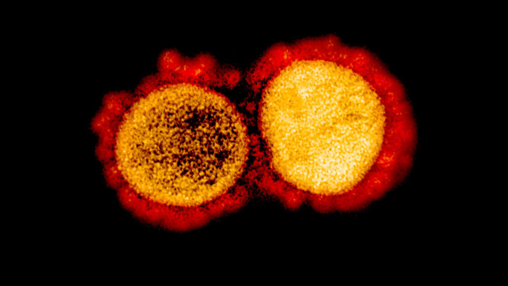
The emergence of the SARS-CoV2 virus has caused turmoil across the globe, forcing the researchers to study it in more detail. However, the behavior of the virus has been quite notorious. The frequent mutations and very less prior knowledge has caused some obstacles. However, scientists were acquainted that if they understand the underlying pathways in lung cells and host protein being impacted during the viral infection then there can be chances of identifying potential solutions. Considering this, scientists have recently found out how SARS-CoV2 hijacks and damages the lung cells.

The study was a multi-group collaboration between the Center for Network Systems Biology (CNSB), Center for Regenerative Medicine (CReM), and National Emerging Infectious Disease Laboratory (NEIDL). The researchers have mapped the molecular responses emerging from lung cells infected with the SAR-CoV2 virus. The finding of the study has been published in the Journal of Molecular Cell.

To understand the disease pathology and gain new insights to discover potential therapeutic targets, the researchers bioengineered the human alveolar cells. The cells when combined with high-end mass spectroscopy technology, it helped the researchers to identify potential host proteins and pathways changing during SARS-CoV2 infection. The researchers found out that lung cells infected with SAR-CoV2 have abnormal phosphorylation.

Phosphorylation is a very crucial protein modification process contributing to protein functionality in the cells. The proper phosphorylation process is important for healthy cells. However, it was noticed that the infection alters this process leading to cascading abnormal changes. These changes increase the chances of the virus to thrive within the cells and eventually destroy it.

The study also showed that as soon as the virus encounters the lung cells, it initiates exploiting the resources required for the normal functioning and growth of the cell. The invasion of the virus further disrupts the functioning of the cell and damaging it extensively. The resources used, powers the virus eventually leading to rapid proliferation and expansion in nearby regions. As a result, the exhausted and damaged cells undergo self-destruction and the virus starts infecting the cells in the vicinity while evading the body’s immune system. This cycle repeats continuously leading to the hijacking of lung cells and widespread damage.

The researchers examined lung alveolar cells from one to 24 hours after infection with SARS-CoV-2 to understand what changes occur in lung cells immediately (at one, three, and six hours after infection by SARS-CoV-2) and what changes occur later (at 24 hours after infection). These changes were then compared to uninfected cells. All proteins from infected and uninfected alveolar cells, corresponding to the different time-points were extracted and labeled with unique barcoding tags called “tandem mass tag.” These tags, which can be accurately detected only by a mass spectrometer, permit robust quantification of protein and phosphorylation abundance in cells.

“Our results showed that in comparison to normal/uninfected lung cells, SARS-CoV-2 infected lung cells showed dramatic changes in the abundance of thousands of proteins and phosphorylation events,” said Darrell Kotton, MD, BUSM, CReM.

“Moreover, our data also showed that the SARS-CoV-2 virus induces a significant number of these changes as early as one-hour post-infection and lays the foundation for a complete hijack of the host lung cells,” adds Elke Mehlberger, PhD, NEIDL.

Scientists also tried to examine the data obtained from the study to identify any potential opportunities for COVID-19 treatment. They found that about more than 18 existing drugs that have been already clinically approved can be re-purposed for the treatment. The research team believes that the current findings are very crucial and can contribute a lot to the field, specifically in terms of devising a cost-effective, robust and life-saving treatment to overcome COVID-19.


Antibiotic Resistance- An ongoing crisis

Antibiotic resistance is an ancient and mounting problem. Common causes are overpopulation, increased use of antibiotics, and even due to enhanced global migration.  For a long time, antibiotic treatment has been the main approach of treatment in modern medicine to combat infections. The golden era of antibiotics was between the 1930s to 1960s, giving rise to many antibiotics. Antibiotic resistance poses a very serious global threat due to the growing concern of human and animal health. This is due to multidrug-resistant bacteria or popularly known as “superbugs”.

The plausible causes of global microbial resistance include overuse of antibiotics in animals consumed as food by humans. Other superficial causes include, increase in international travel and poor hygiene. These factors mainly play a role in the genetic selection in a community of resisting antibiotics. 

History and Benefits of Antibiotics:

The first documented use of antibiotics was with the discovery of penicillin by Alexander Fleming during the world war. Penciliine was successful in controlling the bacteria and was said to have saved millions of lives. Shortly after the boom in usage, the penicillin resistance was a major problem, threatening many of the advances in the medical field at that time. What was thought as just penicillin resistance later proved to be a multidrug resistance. Microorganisms under Darwinian natural selection to develop the resistance. Naturally, most antibiotics are produced using bacteria or environmental fungi, and few are completely synthetic using sulphonamides and fluoroquinolones. 

Not only saving lives, but antibiotics have also played a vital role in advancing major breakthroughs in the field of surgery and medicine. They have successfully prevented and treated infections that commonly occur in patients undergoing chemotherapy, who are also suffering from chronic diabetes or rheumatoid arthritis. 

Overuse:

Even as early as 1945, Alexander Fleming with the discovery of penicillin, also warned the public that the era of antibiotics will also lead to the era of misuse and resistance. Epidemiological studies depict a direct relationship between the consumption of antibiotics and the rise in bacterial resistant diseases. These bacteria can be transmitted or inherited between relatives and friends. Resistance can also occur due to spontaneous mutation. Antibiotics remove the sensitive competitors and leave the resistance bacteria by natural selection to reproduce and multiply more. Despite these warnings, antibiotics are overprescribed throughout the world. 

Agricultural use:

In both developing and developed worlds, antibiotics are seen to be used very frequently as growth supplements in livestock. According to a census, almost 80% of the antibiotics sold throughout the world are used in animals to prevent infection and promote growth. Treating livestock with antimicrobials can improve overall health, produce larger yields, and an overall high-quality product.  

The antibiotics used in treating livestock is in turn consumed by the top of the food chain- humans. This transfer of resistant bacteria to humans was first noticed 35 years ago when the antibiotic rates were found high in the intestines of both farm animals and farmers. Recently molecular detection methods prove that resistant bacteria from farm animals reach humans through consumption. 

It should also be noted that the agricultural use of antibiotics also thrive in urine and stool. These are widely dispersed through fertilizers affecting the groundwater and surface. While this may account for a small fraction of overall antibiotic use, the end geographical spread can be a considerable size. This practice also affects the micro

The emergence of resistance:

Organisms develop resistance by several techniques including altering the target site of binding, inhibiting the drug entry, and enzyme production that leads to the degradation of antimicrobials.Various antimicrobial drugs like antibiotics produced by saprophytic bacteria tend to develop mutual benefits with the other organisms surrounding it and sometimes even inhibit growth. Available data suggest that the sublethal concentration is the antibiotics have a significant impact on the microbial flora, and may even be effective in signalling molecules which may induce a microbial and host gene expression. 

Another important finding reveals that few saprophytic bacteria is capable of producing a broad-spectrum antibiotic known as carbapenems. Various genes involved in constructing this antibiotic may also play an important role in biofilm formation. These findings reveal more unexpected impacts of the antibiotics.

More knowledge is needed to the extent of the broad-spectrum antimicrobial resistance. Current panic is due to the inadequate information. The future cannot be predicted with surety at the stage with regards to the resistance with the unavailability of novel antibiotics. Multiple well thought strategies need to be in place to confront this particular issue. Regulations should be implemented by every country to monitor prescriptions and the use of antibiotics. Environmental and ecological issues should not be ignored and all elements should be part of the control policy. Alternatives to antibiotics such as lytic bacteriophages vectors and probiotics can potentially help to decrease the use of antibiotics. 

Use of Computational Methods in Stem Cell Biology

Use of Computational Methods in Stem Cell Biology

For a few decades now, the field of developmental biology has utilized computational technologies to explore the mechanisms of the developmental process. It was first in the 1950’s Alan Turing wrote a computer program that was able to model how morphogen concentrations can affect the growth of an in vitro embryo. Since then several techniques have been developed that can generate comprehensive data of a molecular type also known as OMICs. 

Though the use of computational methods was largely limited to the theoretical mechanisms, the birth of large genome sequencing, paved the way to process large molecular data. Computational models complement the statistical data by providing mechanistic insights into the biological processes and by the ability to predict future outcomes in terms of biological processes that can guide experimental research. 

Difference between Mechanistic models and Machine Learning Models:

For several years, Machine Learning (ML) approaches have been used for pattern recognition, prediction, and classification of biological systems, especially system cell research. Some of the important examples include the construction of 3D stem cell images from fluorescent microscopic results. Ml can also predict the experimental conditions and determine future outcomes.

Although ML has a decent accurate predictive power, they require large amounts of data especially imaging and omics datasets for interpreting statistical relationships between the input and predicted output data. ML usually specializes in predicting the outcome but not revealing the underlying complex processes, preventing them from providing any mechanistic insights on the biological processes. ML can be classified as supervised and unsupervised learning. The supervised learning can predict outcomes of foreseen data by studying the labeled training data, whereas unsupervised tries to make sense of any unlabelled data by extracting in-depth features and patterns of its own. 

By contrast, mechanistic models generally rely on the mechanistic hypothesis implied from the experimental data to predict novel outcomes and describe the behaviors of the whole system. These models are often assembled based on the simplified mathematical and conceptual formulations of the observed experiment. Moreover, a single based cell experiment of this model has been developed to elucidate cell fate dysregulation linked with congenital diseases. 

Applications of Computational Methods in Regenerative Medicine

It is well known that cell transplantation especially using induced pluripotent stem cells (iPSCs) is one of the main strategies in regenerative medicine to reinstate damaged or ill-functioning cells. Though various clinical applications using iPSCs are underway there are still few challenges that need to be overcome before it reaches its full potential. One of the main ways to overcome this problem is by figuring out the in-vitro manufacturing of the donor cells to gain appropriate knowledge of the cell expression- the identity of host tissue cells. 

Current techniques have a low conversion efficiency, forcing the researchers to spend a large number of their resources in order to get an accurate result. Moreover, cell conversion often results in creating unnecessary immature cells or non-variants of the cells, ultimately failing to reciprocate the desired functionalities and phenotypes. On the other hand, computational methods can help in achieving the desired results. The latest advancements in scRNA sequencing technologies can help the researchers to accurately characterize functionally gene expression and cell subtypes. 

By combining the computational methods with existing novel experimental techniques, it is possible for researchers to now open up to new avenues in designing protocols and treatments for congenital disorders and for enhancing regeneration of cells. 

Stem cell rejuvenation is another strategy promising to prevent the damaged stem cell function and to help optimize tissue repair processes in age-related or degenerative disorders. The main reason behind impaired stem cell function is the disruption of pathways of the endogenous stem cells due to certain mutations or aged niche. The computational models can help in determining this particular impaired niches and signaling pathways and further help in proving insights with the mechanisms of the cell dysregulation in aging. Researches can use these predicted signaling molecules to counteract a niche effect for rejuvenating stem cells.  

Future Perspectives:

As discussed a number of challenges in the research can be resolved with the development of multiscale computational methods. With the increasing work in single-cell expansion and scRNA data, it is now possible to develop complex computational methods, including cell-cell communication and intracellular network-based models. 

Although ML has been employed successfully in pattern recognition and classification, they are not capable of providing information on biological processes. The implementation of mechanistic models with ML can help in a better understanding of mechanisms and predictions based on simple assumptions. In the future, stem cell researchers could coordinate with computational models, before performing an experiment to address certain biological questions and assess the required data for the model.

Use of nanoparticles in drug carrier systems

Since ancient times, plant-based natural products have been commonly used as medicines. Nearly as much as 25% of major pharmaceutical compounds are derived from natural resources. They exhibit fascinating chemical diversity and biological properties with minimal toxicity.

Despite the advantages, companies are hesitant to invest more in natural delivery systems, due to concerns such as size, shape, and biocompatibility. The use of large materials in drug delivery includes many risks such as solubility and poor absorption. Such problems gave rise to nanotechnology, currently revolutionizing the field of medicine. 

Nanotechnology is an advanced field of nanoparticles being used in the science field at a molecular level. Nanoparticles are as small as 10nm to 1000nm, usually preferred to be less than 200nm. This helps them to move freely inside an organism when compared to other materials. Lately, the use of nanoparticles in drug delivery systems is speculating a lot of discussions.

Nanoparticles in drug delivery:

Currently, one of the challenges faced by researchers is to deliver a drug to an accurate site without causing any potential side effects to other healthy organs. This is especially important for cancer treatment, where the tumor is a distant metastasis in various organs. Nanoparticles employed, have specific properties to overcome the above limitation. These include their small size, ability to penetrate cell membranes, binding and stabilizing proteins. Nanoparticle entrapment of drugs is performed to make sure the drug is delivered to the target tissue supported with a controlled release. 

The idea of using nanoparticles with natural compounds is very attractive in recent times. Natural compounds have a lot of properties that are enhanced by combining with nanoparticles. These properties include targeting a specific tissue and controlled release of the drug. 

Metallic, inorganic, and polymeric nanoparticles are some of the frequently used types in performing a target-specific drug delivery system. In particular, drugs with poor solubility need the aid of these nanoparticles. 

The efficacy of the use of nanoparticles varies according to the material, size, and other biodegradability properties. Due to their biocompatibility properties, various synthetic polymers such as polyvinyl alcohol and poly-L-lactic acid, and natural polymers such as chitosan and alginate are broadly used in fabricating nanoparticles. 

Polymeric nanoparticles can be divided into nanocapsules and nanospheres, both of which are proven to be attractive drug delivery systems. The use of an optimal nano-drug delivery system is based upon the properties of the drugs chosen for the treatment. 

Possible hazards:

When combining nanotechnology in medicine, toxicity exhibited by nanoparticles cannot be ignored. For pharmaceuticals, specific drug formulations can be put in place for clinical efficacy and minimizing toxicity. 

Nanoparticles are known for their unique surface properties, and since it is the contact layer with the body tissue, it should be evaluated from a toxicology standpoint. Although a lot of tests and procedures are available to evaluate the material, it cannot be assumed that these tests will be precise in detecting all risks. This will most likely depend on the origin of the materials- biological or non-biological.

Recently, researchers are exploring the ideas of combining nanoparticles and natural products to minimize toxicity. Both individually have already been in use for several years, but have their own limitations. The greenway of formulating nanoparticles is widely encouraged as it lowers the hazardous constituents during the synthesis process. By using green nanoparticles, the chances of side effects can be minimized. Furthermore, by changing the shape, size, and hydrophobicity we can further enhance the bioactivity of nanoparticles.

Future of nanoparticles in the medicinal field:

Nanoparticles in the medicinal field is currently one of the fascinating areas of research out there. In the future, by the looks of it, the field of cancer looks to benefit the most from nanoparticles. By using various types of nanoparticles, it is possible to transport an accurate amount of drug to the tumor cells without causing harm to other cells. The application of nanoparticles in medicine will definitely be the future trend in diagnosis and treatment research. 

More research needs to be performed to achieve a more consistent drug loading and release capacity. , progress is being made in the use of metal-based nanoparticles like gold and silver in diagnosis and therapy areas of research. 

Nanotechnology is truly a multidisciplinary science beneficial to drug delivery systems. Despite the overwhelming advantages, its actual impact on the healthcare system is quite limited. In the future, a more conceptual understanding of biological response to nanoparticles must also be studied. Ultimately researchers should be able to deliver drugs for a long period of time with great precision and controlled release.

Small interfering RNA (siRNA) Technology

Small interfering RNA (siRNA) Technology

In the past two decades, there has been increasing awareness of the role RNAs play in the regulation of gene expression. The field of RNA was revolutionized with the discovery of RNA interference (RNAi).

RNAi is a regulatory mechanism seen in most of the eukaryotic cells that contain double-stranded RNA (dsRNA) which in turn triggers direct homology dependent control of gene activity. Popularly known as small interference RNAs (siRNA), dsRNA is usually 21–22 bp long and has characteristics 2 nt 3’ overhangs which helps them to be recognized by the mechanism of RNAi. 

Targeting and manipulating RNAi pathways can be a potential tool to change any biological process post-transcriptionally in many health conditions including g  autoimmune diseases and cancer among others. RNAi was described as “Breakthrough of the year” by the journal Science in the year 2002, having the potential to be a powerful drug for several diseases. Optimum designing of siRNA can enhance the stability and specificity of the RNAi process and prevent off-target effects. 

Design:

When considering siRNAs for therapeutics, it is essential for the selection of appropriate designs that have good potency, stability, and specificity. Over the years, protocols have been put in place to assist in designing an ideal siRNA, based on the target of interest. Once synthesized these siRNAs should be experimentally evaluated to determine its effects in gene silencing. Though tested to be effective, at times siRNAs have off-target effects, where unrelated genes tend to be altered. Furthermore, siRNAs may also induce an unnecessary innate immune response which could potentially initiate a harmful inflammatory response in patients. 

Stability:

When using siRNA technology for therapeutics in in-vivo models, the stability of the molecule plays an important factor. When unprotected, siRNA is highly unstable and can easily be degraded in the human plasma, with very minimal half-life. At times like these, siRNA is chemically modified to increase the half-life without altering the efficacy. Such chemical modifications usually happen in the phosphate group of the molecules hat confers exonuclease resistance or alterations in the sugar residues that confer endonuclease resistance. A right balance between increased stability and efficacy is the right way to configure a siRNA for in-vivo use.

Delivery:

It is safe to say that no matter the efficacy or stability it is most challenging to deliver the right siRNA to the targeted tissues/ cells. Due to its negative charge, siRNA molecules are incapable of penetrating cell membranes easily. For this reason, many strategies have been developed like lipid-based formulations. Most of these strategies are non-specific and are build into delivery agents. 

Recently, study results showed that single-stranded siRNA is capable of performing gene silencing in-vivo. This finding is particularly significant since single-stranded siRNA has multiple advantages over double-stranded siRNA. The lack of second passenger strands makes it more potent and reduces the risk of uncalled alterations. 

Applications in medicinal fields:

Due to the effective gene silencing, siRNA is popular among medicinal fields. The degree of specificity is also an added advantage, as is its ability to utilize numerous RNA sequences to target specific cells and deliver drugs or medicines. The ultimate goal for scientists is to find an optimal way to kill or slow down the disease without affecting the surrounding tissues. In addition, the ability to specifically target genes that cause cellular damage or destruction enables pharmaceutical experts to specifically target those genes by the use of siRNA interference. 

The progression from the initial discovery of siRNA technology to clinical applications has been groundbreaking. Understanding the fundamentals behind the technology has led to its widespread application in both basic research and clinical applications of treating a disease. 

Despite the significant progress made in the field of RNAi technology, there is still a lot of more research that needs to be conducted to perfect the design Before siRNA becomes a common commercial therapeutic technology in the clinical field, a number of major hurdles need to be addressed. Lately, the difficulties associated with inefficient delivery to target cells have been brought into the light. There are also problems associated with the reduced biostability of unmodified RNA. 

Regardless of certain setbacks, the hope still remains that with the time, siRNA technology will prove to be the next “Superdrug” against many destructive diseases. In addition, RNAi has proven to be an important tool and has opened a new world of basic investigation. 

Source

Exploring CAR-T-cell therapy using CRISPR technology

Exploring CAR-T-cell therapy using CRISPR technology

Immunotherapy is the lesser-known mainstream treatment for cancer. It has recently been gaining popularity ever since the first chimeric antigen receptor T- (CAR-T) cell therapy was approved for Non-Hodgkin lymphoma in 2017. Currently, numerous CAR-T-cell therapies for a variety of cancers are being granted investigational new drug clearance to enter clinical phases.

The clustered short palindromic repeat or also known as CRISPR associated protein 9 (CRISPR/Cas9) technology plays a crucial role in advancing the CAR-T-cell therapy field, owing to its high efficiency, simplicity, and flexibility. It is an exciting new world out there for CAR-T cell therapy researchers, aiming to term cancer a curable disease. 

What is chimeric antigen receptor T- (CAR-T) cell therapy?

CAR-T cell therapy usually involves engineered T cells that act as synthetic receptors. They typically contain a tumor-specific chimeric antigen (CAR) containing an intracellular domain, an antibody derived targeting extracellular domain and transmembrane domain. The transmembrane domain from CD28 is responsible for providing stability to CAR. 

The T cells programmed with CARs can be used to specifically target and kill antigen-expressing cells without the major histocompatibility complex. Data from studies show that CAR-T-cell therapy has helped in the complete remission of patients diagnosed with a variety of solid and hematologic cancers, especially in relapsing cases of acute lymphoblastic leukemia with a remission rate of 80-100%. 

CRISPR technology in developing CAR-T-therapy:

One of the crucial decisions in designing CAR-T cells is choosing the right DNA template for CAR expression. An appropriate DNA template should be obtained easily and rapidly, containing flexible insert sizes, highly efficient target sites, and low cellular toxicity. For a long time, viral vectors were used, but concerns regarding the integration in the wrong location causing unnecessary diseases, gave rise to CRISPR/Cas9 technology. 

A powerful eukaryotic cell genome editing tool, CRISPR/Cas9 technology makes it possible to insert large genes at the required genetic sites in T cells for successful CAR-T engineering without viral vectors. The two essential components of CRISPR technology include a guide RNA (gRNA) customized to recognize the protospacer on target DNA, and a Cas9 protein to create precise double-stranded breaks (DSBs) for gene mutation. 

DSBs have a unique ability to create two distinct mechanisms for repair. One is through the non-homologous joining, which introduces mutations to DSB sites and the other a homology-directed repair (HDR) mechanism which makes sure the donor DNA template is accurately placed for the gene knock-in. The HDR mechanism is popular among researchers due to its precision insert of the CAR expression cassette into the T cells. 

Methods employed to prevent allogeneic CAR-T side effects:

The multigene editing capability of CRISPR/Cas9 technology is employed for the potential safety of any allogeneic CAR-T therapy-associated side effects. For example, to prevent any graft Vs host rejection, the general approach would be to knock out the expression- TCR-αβ of T cells. To function, TCR-αβ requires both α- and β-chains. The α-chains encoding TCR-α can be knocked out using CRISPR gRNA. From previous studies, it can be noticed that when CAR placed under endonuclease transcriptional regulation, it leads to continued T cell function and a delay in cell exhaustion. 

Host Vs graft disease can also be avoided by knocking out β2-microglobulin, an essential part of the major histocompatibility complex class I molecules, using CRISPR to stop the surface antigen presentation. The gRNAs have also been shown to target immune inhibitory receptors enhancing the antitumor activity of CAR-T-cells. 

Future Perspective:

The remarkable use of CAR-T cells in the remission of advanced malignancies is promoting the rapid growth in developing smarter and commercialized CAR-T therapies. The CRISPR/Cas9 genome editing technology promises a hopeful next-generation CAR-T cell product by adding novel CAR-T cells knockout and inducible safe switches to avoid self-killing. 

However, there are certain concerns regarding the technology which includes off-target effects, causing random mutations. Multiple strategies such as optimized gRNA design, careful selection of target sites, prior off-target detection assays should be attempted to minimize the risks. 

By technical progress to avoid mutations, and improved delivery efficiency, CRISPR/Cas9 mediated T cell engineering holds great promise. Currently, experts are working on exploring other CRISPR/Cas 9 gene targets for multiplex editing for potentially developing an optimal off-shelf allogeneic CAR-T cells products as universal treatment options.

Source

Versatile use of Bacterial nanocellulose for wound healing applications

Versatile use of Bacterial nanocellulose for wound healing applications

Over the years, several therapeutic options have been available for wound and burn treatment. The urgent need for better strategies to accelerate treatment leaves more scope for therapeutic improvement in this field. 

Cellulose is one of the most naturally occurring polymers from renewable sources. Occurring in the form of a linear homo-polysaccharide it consists of β‐d‐glucopyranose units linked by β‐1,4 glycosidic bonds. In modern times, bacteria is one of the commonly used sources for producing cellulose also known as bacterial cellulose. Recently, experts have been playing around with the idea of bacterial Nanocellulose (BNC), cellulose constructed using nano-engineering. 

Bacterial nanocellulose matrix has outstanding mechanical and physical properties courtesy of its unique 3D structure. BNC aggregates to form long fibrils, providing room for high elasticity, surface area, and resistance. Such intrinsic characteristics make it the best choice for wound dressings or protecting injured tissues. It does help that BNC is also non-carcinogenic, non-toxic, and biocompatible. 

Bacterial nanocellulose in wound healing:

It is well known that the largest organ of the body is skin. In its native state, the skin is usually dry and acidic in nature. Altered skin integrity is usually caused by systemic factors such as nutrition, among others. When an individual suffers from serious skin damage due to an accident or disease, a complex series of the biological processes are involved in restoring the lost skin. 

A perfect wound dressing must be able to retain moisture and allow oxygen exchange accelerating healing time and preventing infection. Experts consider BNC to be one of the most suitable materials for wound treatments due to its characteristics such as favorable mechanical properties, chemical purity, and water-absorbing capacity. BNC in its natural state has consistently shown great capacity to stimulate wound healing. To further improve the healing effect of BNC, the material can be combined with natural additives such as proteins, glycosides among others, to improve the mechanical strength and cellulose-based dressings with antimicrobial properties. 

Incorporating mesenchymal stem cells into bacterial nanocellulose:

One of the recent strategies to improve BNC wound dressing is the incorporation of mesenchymal stem cells (MSC) in the matrix. MSCs are adult pluripotent stem cells that are expected to integrate into the victim’s tissue and promote regeneration of the damaged tissue.

Several studies prove that MSC can evolve into various cell types including muscle bone and cartilage. They have a great capacity to self-renew while maintaining its integrity, an essential feature needed to improve the wound healing process, and inducing re-epithelization of the wound. 

Genetically engineered bacterial nano cellulose:

Genetic engineering of the BNC is currently being explored with an aim to optimize the properties of the matrix and the cost-effectiveness of the manufacturing process. Previously, strain improvement was performed through the transfer of BNC related gene determinants to a previously prepared “cell factory” organism. This was done to produce a heterologous expression of genes.

Recently a study used a small RNA (sRNA) interference system to improve the native cellulose production path. The constitutive production of the BNC was shut off to prevent any mutants, a common phenomenon in a well-aerated environment. This was replaced by expression vectors to functionalize BNC with specific proteins, by fusing the genes encoding the protein of interest to the short nanocellulose binding domains. 

Challenges and Future Directions:

Using nano engineering in the field of tissue engineering has opened up a lot of new prospects. Experts are looking to develop BNC based out of commercial 3D printing materials, as an alternative to the chemical products such as resins, synthetic thickeners, and plastics, Another added advantage of 3D printed BNC is the possibility of creating flexible and adjustable dressings. The option of 3D printed nanofiber based bacterial cellulose can also offer an opportunity to develop wearable biomedical devices as sensors and drug-releasing materials, to monitor the patient’s wounds constantly. 

Another interesting discovery on the works is the creation of transparent wound coverings using nanocellulose. This discovery will allow optical real-time monitoring of wound healing and help in diagnostics of viral infections and inflammations in chronic wounds. 

In conclusion, although BNC has made substantial progress in the field of tissue engineering, one of the common drawbacks is the non-degradability of nanocellulose in human organisms. This could potentially lead to scar formations and other complications when intended for direct use. However, artificially constructed skin can be used for experimental studies, such as metabolism, and vascularization of skin tissue.

Currently, there are no materials yet to be found that can fully capture the intricates of the native tissue to restore function at an ideal level. The remaining challenge will be to innovate new composite materials using nanoscale engineering to produce fully biomimetic tissues. As the complexity of applications increase, an active remodeling of the existing scaffolds will be required. 

Source

A brief insight on ACE receptors role in COVID-19

A brief insight on ACE receptors role in COVID-19

With the novel coronavirus, COVID-19 is spreading across the world and the fact that no drug or treatment has been found against it is creating fear among people. Coronavirus is a large family of enveloped, single-stranded RNA that infects mainly mammals including humans. In humans, coronavirus causes mild to severe respiratory illness. These viruses exhibit strong virulence and are highly contagious. While a person infected, produces mild symptoms, certain individuals respond severely, sometimes leading to death. 

The SARS pandemic, back in 2002 is known to belong to the same family of viruses as COVID-19. According to WHO, SARS rapidly spread through 29 countries, with 8096 confirmed cases, but the current pandemic has surpassed all numbers by infecting over millions of people across the world. Today, it is due to SARS that has resulted in a coordinated effort to develop treatments targeting the virus or host cell components responsible for viral replication.

The deadly virus, COVID-19 enters the human body and binds to the target cells through angiotensin-converting enzyme 2 (ACE2) which is mainly expressed in endothelial cells and Leydig cells. ACE-2, a transmembrane metallo carboxypeptidase, is an enzyme which for years has been important for the treatment of hypertension. With further Polymerase Chain Reaction (PCR) analysis it was determined that the ACE-2 receptor is also present in the lung and gastrointestinal tract, tissues harboring COVID-19. 

Inhibiting ACE2 receptor blocks COVID-19 entry:

ACE-2 belonging to the family of ACE receptors, plays a key role in the Renin-Angiotensin System (RAS) and in the treatment of hypertension. ACE2 is known to degrade angiotensin II and thereby, negatively regulating RAS. Recently experts have revealed that the COVID-19 virus uses ACE-2 receptors as their entry in HeLa cells. Additionally, it was found that by using anti- ACE-2 antibodies in other mammals like monkeys, it was seen that there was an entry blockage of pseudotypes expressing the COVID-19 virus. 

For the virus to enter the host cell, its spike glycoprotein (S) needs to be cleaved at 2 sites, termed S protein primming so the viral and host cells membrane can fuse. A serine protease TMPRSS2 is essential for cleaving the S protein. It was found by treating Calu-3 human lung cell line with a serine inhibitor- camostat mesylate the entry of the COVID-19 virus was partially blocked. Angiotensin-converting enzyme (ACE) inhibitors is also found to play a role in preventing the formation of angiotensin II. ACE multifaceted functions include treating heart failure, controlling high blood pressure, and preventing kidney failure in diabetic patients. 

Existing concerns about ACE inhibitors:

Though ACE inhibitors might seem like a promising solution for COVID-19, there are certain concerns regarding the increased susceptibility to COVID-19. These are considered based on the fact that ACE inhibitors are also used in treating millions of people with hypertension, heart, and kidney disease. When administered with inhibitors, diabetes and hypertension patients were observed to have an increase in ACE2, which in turn would facilitate infection with COVID-19. It is hence hypothesized that treating diabetic and hypertension patients with ACE inhibitors might make them more susceptible to COVID-19. 

Furthermore, several studies have reported that long term usage of ACE inhibitors can modify the adaptive immune response, which is a key and much-needed defense against any infection. These particular effects must be taken into noticed and investigated further in context to COVID-19. 

Road to COVID-19 therapies:

These findings could greatly impact the efforts being taken in developing treatments for the current pandemic. For instance, TMPRSS2 inhibitors can be potentially used to prevent virus entry into the host cells. Though there are certain drawbacks in using ACE inhibitors, there is a lack of scientific evidence and clinical data to support the discontinuing of ACE inhibitors in COVID-19 patients with existing hypertension and diabetes. The proof that reduction in mortality due to ACE inhibitors and the beneficial effects outweigh the theoretical risks. 

Our interpretation of this hypothesis should not lead to changing drugs for patients with diabetes or hypertension, without consulting an expert physician. Though it’s of utmost urgency for the scientific community to come up with some solution for this deadly virus, further research and clinical trials need to be performed before any of the said theory can become a reality. 

Source

Mutant Polio Virus from Vaccine more infectious than the wild type

Mutant Poliovirus from vaccine more infectious than the wild type

How it all began?

Back in the 20th century, there were many more diseases that worried parents then Polio did. Polio struck during summer, making its way through towns, every few years. Most people were reported to recover quickly, though some suffered temporary or permanent damage leading to paralyzation and even worse, death. With many polio survivors disabled for life, it was a constant reminder to the society the toll it took on young lives. Polio reached the level of epidemic proportions back in the 1900s affecting the infant and young kids, where the infant’s immune system is still aided by maternal antibodies could not fight the virus. 

Polio is caused by a family of viruses belonging to the Enterovirus genus. These viruses are highly contagious usually spreading through contact with people either by oral or nasal secretions, or by contacting a contaminated area. The virus enters the mouth and is found to multiply by the time it reaches the digestive tract, where it continues to multiply. In cases of paralytic polio, the virus makes its way from the digestive tract to the bloodstream attacking nerve cells. 

Development of vaccines to eradicate polio: 

A vaccine is made from a very small amount of wither weak or dead germs that cause the disease, for example-poliovirus. When administered, the vaccine introduces the said virus into the body to trigger an immune response, causing it to recognize and combat the actual disease if encountered in the future. 

 The antibodies specific to the poliovirus was first discovered in 1910. Using immunologic techniques, it was in 1931 different serotypes of the poliovirus were identified. By identifying that the virus can be grown in large amounts using tissue culture, it was just a short time before the first inactivated polio vaccine (IPV) was developed. With human trials being deemed a success, the number of polio cases drastically dropped over the years. Later an improved version of the vaccine using live attenuated virus was developed, which could be administered orally, known as oral poliovirus vaccine (OPV). Soon it became the predominant go-to vaccine for developing countries all over the world, declining the number of cases drastically till a new problem raised recently. 

Vaccine-derived poliovirus:

Over the past few years, more than 10 billion doses of OPV have been administered to millions of children worldwide, preventing more than 10 million cases during that period, bringing down the number of cases drastically. 

So far in the year 2017, only as many as 6 “wild” polio cases were detected worldwide. By wild it means that the number of cases affected by polio “wild type” strain found naturally in the environment. Recently, new cases have been reported of children paralyzed by a vaccine-derived poliovirus. Around nine new cases were identified in countries- Nigeria, the Democratic Republic of the Congo, Central African Republic, and Angola last November, along with Afghanistan and Pakistan. The WHO reported that as long as a single child is infected, all the other children are at risk. 

In developing countries, the oral vaccine is used profusely among all children due to its low cost and accessibility. The vaccine-associated paralytic polio is caused by a strain of poliovirus which was previously termed wild type. The onset was found to be caused by a type 2 virus contained in the oral vaccine. Type 2 virus was a wild type virus eradicated years ago, but in rare cases can mutate into a form that can breach the vaccine protection. 

Erradication possible:

With the COVID-19 outbreak, the WHO has also additionally undertaken the goal of eradicating the new mutant poliovirus before its too late. The role model here is smallpox, which was completely wiped out thanks to a consistent vaccination strategy. The same applies to polio. Similar to smallpox, the polio vaccine also offers impeccable protection, though not applicable to virus mutants. Reports state that the latest mutant outbreak is due to the low vaccination rates. The rise in vaccine-derived polio cases is due to the mutant form of the disease affecting the non-vaccinated children through contaminated matter. 

The coverage of vaccination and hygiene measures must be extended so that the new mutant can no longer continue to survive, the same way the previous polio epidemic in Congo was eradicated. Though the current vaccines appear to be good enough to be effective, the new pathogen is nonetheless a warning. The need for new protectant vaccines is more important now than ever. It is only this way there is a chance of permanently wiping out polio. 

Source