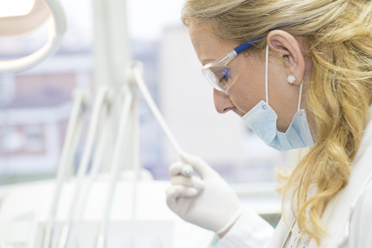
The emergence of the SARS-CoV2 virus has caused turmoil across the globe, forcing the researchers to study it in more detail. However, the behavior of the virus has been quite notorious. The frequent mutations and very less prior knowledge has caused some obstacles. However, scientists were acquainted that if they understand the underlying pathways in lung cells and host protein being impacted during the viral infection then there can be chances of identifying potential solutions. Considering this, scientists have recently found out how SARS-CoV2 hijacks and damages the lung cells.
The study was a multi-group collaboration between the Center for Network Systems Biology (CNSB), Center for Regenerative Medicine (CReM), and National Emerging Infectious Disease Laboratory (NEIDL). The researchers have mapped the molecular responses emerging from lung cells infected with the SAR-CoV2 virus. The finding of the study has been published in the Journal of Molecular Cell.
To understand the disease pathology and gain new insights to discover potential therapeutic targets, the researchers bioengineered the human alveolar cells. The cells when combined with high-end mass spectroscopy technology, it helped the researchers to identify potential host proteins and pathways changing during SARS-CoV2 infection. The researchers found out that lung cells infected with SAR-CoV2 have abnormal phosphorylation.
Phosphorylation is a very crucial protein modification process contributing to protein functionality in the cells. The proper phosphorylation process is important for healthy cells. However, it was noticed that the infection alters this process leading to cascading abnormal changes. These changes increase the chances of the virus to thrive within the cells and eventually destroy it.
The study also showed that as soon as the virus encounters the lung cells, it initiates exploiting the resources required for the normal functioning and growth of the cell. The invasion of the virus further disrupts the functioning of the cell and damaging it extensively. The resources used, powers the virus eventually leading to rapid proliferation and expansion in nearby regions. As a result, the exhausted and damaged cells undergo self-destruction and the virus starts infecting the cells in the vicinity while evading the body’s immune system. This cycle repeats continuously leading to the hijacking of lung cells and widespread damage.
The researchers examined lung alveolar cells from one to 24 hours after infection with SARS-CoV-2 to understand what changes occur in lung cells immediately (at one, three, and six hours after infection by SARS-CoV-2) and what changes occur later (at 24 hours after infection). These changes were then compared to uninfected cells. All proteins from infected and uninfected alveolar cells, corresponding to the different time-points were extracted and labeled with unique barcoding tags called “tandem mass tag.” These tags, which can be accurately detected only by a mass spectrometer, permit robust quantification of protein and phosphorylation abundance in cells.
“Our results showed that in comparison to normal/uninfected lung cells, SARS-CoV-2 infected lung cells showed dramatic changes in the abundance of thousands of proteins and phosphorylation events,” said Darrell Kotton, MD, BUSM, CReM.
“Moreover, our data also showed that the SARS-CoV-2 virus induces a significant number of these changes as early as one-hour post-infection and lays the foundation for a complete hijack of the host lung cells,” adds Elke Mehlberger, PhD, NEIDL.
Scientists also tried to examine the data obtained from the study to identify any potential opportunities for COVID-19 treatment. They found that about more than 18 existing drugs that have been already clinically approved can be re-purposed for the treatment. The research team believes that the current findings are very crucial and can contribute a lot to the field, specifically in terms of devising a cost-effective, robust and life-saving treatment to overcome COVID-19.