Use of Computational Methods in Stem Cell Biology

Use of Computational Methods in Stem Cell Biology

For a few decades now, the field of developmental biology has utilized computational technologies to explore the mechanisms of the developmental process. It was first in the 1950’s Alan Turing wrote a computer program that was able to model how morphogen concentrations can affect the growth of an in vitro embryo. Since then several techniques have been developed that can generate comprehensive data of a molecular type also known as OMICs. 

Though the use of computational methods was largely limited to the theoretical mechanisms, the birth of large genome sequencing, paved the way to process large molecular data. Computational models complement the statistical data by providing mechanistic insights into the biological processes and by the ability to predict future outcomes in terms of biological processes that can guide experimental research. 

Difference between Mechanistic models and Machine Learning Models:

For several years, Machine Learning (ML) approaches have been used for pattern recognition, prediction, and classification of biological systems, especially system cell research. Some of the important examples include the construction of 3D stem cell images from fluorescent microscopic results. Ml can also predict the experimental conditions and determine future outcomes.

Although ML has a decent accurate predictive power, they require large amounts of data especially imaging and omics datasets for interpreting statistical relationships between the input and predicted output data. ML usually specializes in predicting the outcome but not revealing the underlying complex processes, preventing them from providing any mechanistic insights on the biological processes. ML can be classified as supervised and unsupervised learning. The supervised learning can predict outcomes of foreseen data by studying the labeled training data, whereas unsupervised tries to make sense of any unlabelled data by extracting in-depth features and patterns of its own. 

By contrast, mechanistic models generally rely on the mechanistic hypothesis implied from the experimental data to predict novel outcomes and describe the behaviors of the whole system. These models are often assembled based on the simplified mathematical and conceptual formulations of the observed experiment. Moreover, a single based cell experiment of this model has been developed to elucidate cell fate dysregulation linked with congenital diseases. 

Applications of Computational Methods in Regenerative Medicine

It is well known that cell transplantation especially using induced pluripotent stem cells (iPSCs) is one of the main strategies in regenerative medicine to reinstate damaged or ill-functioning cells. Though various clinical applications using iPSCs are underway there are still few challenges that need to be overcome before it reaches its full potential. One of the main ways to overcome this problem is by figuring out the in-vitro manufacturing of the donor cells to gain appropriate knowledge of the cell expression- the identity of host tissue cells. 

Current techniques have a low conversion efficiency, forcing the researchers to spend a large number of their resources in order to get an accurate result. Moreover, cell conversion often results in creating unnecessary immature cells or non-variants of the cells, ultimately failing to reciprocate the desired functionalities and phenotypes. On the other hand, computational methods can help in achieving the desired results. The latest advancements in scRNA sequencing technologies can help the researchers to accurately characterize functionally gene expression and cell subtypes. 

By combining the computational methods with existing novel experimental techniques, it is possible for researchers to now open up to new avenues in designing protocols and treatments for congenital disorders and for enhancing regeneration of cells. 

Stem cell rejuvenation is another strategy promising to prevent the damaged stem cell function and to help optimize tissue repair processes in age-related or degenerative disorders. The main reason behind impaired stem cell function is the disruption of pathways of the endogenous stem cells due to certain mutations or aged niche. The computational models can help in determining this particular impaired niches and signaling pathways and further help in proving insights with the mechanisms of the cell dysregulation in aging. Researches can use these predicted signaling molecules to counteract a niche effect for rejuvenating stem cells.  

Future Perspectives:

As discussed a number of challenges in the research can be resolved with the development of multiscale computational methods. With the increasing work in single-cell expansion and scRNA data, it is now possible to develop complex computational methods, including cell-cell communication and intracellular network-based models. 

Although ML has been employed successfully in pattern recognition and classification, they are not capable of providing information on biological processes. The implementation of mechanistic models with ML can help in a better understanding of mechanisms and predictions based on simple assumptions. In the future, stem cell researchers could coordinate with computational models, before performing an experiment to address certain biological questions and assess the required data for the model.

Serum- and glucocorticoid- inducible kinase 2, SGK2, is a novel autophagy regulator and modulates platinum drugs response in cancer cells

Serum- and glucocorticoid- inducible kinase 2, SGK2, is a novel autophagy regulator and modulates platinum drugs response in cancer cells

Many cases of ovarian cancers arise from the epithelial cells of the ovary and fallopian tube. The epithelial ovarian cancer is not a single entity disease but rather are several subtypes, each with its distinct genetic and biological backgrounds. This diversity determines the clinical outcome of the disease, where the patients respond differently to the same treatment and sometimes even different prognosis. 

For the past three decades, the standard treatment for advanced epithelial ovarian cancer is chemotherapy, commonly used platinum-based drugs (PT). Though the majority of the patients achieve complete remission, there are cases who might experience recurrence due to acquired resistance to platinum-based drugs. Cellular diversity in tumors and the microenvironment can lead to chemoresistance. Overcoming PT resistance is one of the major challenges faced in ovarian cancer research. 

Over the years, many experiments have been conducted to identify the particular genes responsible for the mechanism directly associated with the PT resistance. PT resistance is linked to several alterations such as drug inactivation, transport, DNA repair, and apoptosis. Among the general pathways, researchers have also observed autophagy has shown to confer with the metabolic plasticity which is necessary to grow and survive in therapy-induced stress. 

Autophagy is a dynamic catabolic process that aids in the formation of double-membrane vesicles also known as autophagosomes. They help in engulfing cellular proteins and organelles and deliver them to the lysosome. When  autophagosomes merge with lysosomes, the contents are degraded and help in fueling the metabolic pathways. 

In this study, the authors began a mission to find the genes responsible for the PT- chemoresistance. A loss of function screening was performed and unveiled that serum- glucocorticoid- kinase 2 (SGK2) as a novel modulator of platinum-based drug sensitivity. SGK family constitutes three isoforms: SGK1, SGK2, and SGK3. Most of the studies revolving around the SGK family, mention its role in the development of human diseases in cancer and in cellular physiology. SGKs were initially identified as regulators, and pumps in the context of epithelial cells ion transport. The study further demonstrates the previously unrecognized role of SGK2 in platinum based drug sensitivity exerted by the autophagic reflux. 

Starting from identifying SGK2 as a druggable modulator, the study characterized the role of autophagy as an escaping strategy activated by the cancer cells to resist PT treatment. The study later demonstrated that pharmacological or genetic inhibition of the isotype SGK2 could potentially block the autophagic process stimulated by the PT treatment. This evidence throws light on the possibility of developing new anticancer strategies on drug repositioning. Overall the study proves that SGK2 kinase controls the PT induced cell death in epithelial ovarian cancer by inhibiting autophagy.

Isolation of potent SARS-CoV-2 neutralizing antibodies and protection from disease in a small animal model

Isolation of potent SARS-CoV-2 neutralizing antibodies and protection from disease in a small animal model

For the past few months, there has been a global spread and toll of COVID-19. So far, humanity has been able to eradicate only one other human infectious disease- smallpox. The novel infectious disease- COVID-19 has had its devastating share of lives globally and currently there is no cure or licensed vaccine. 

Many studies lately have been discussing in-depth about neutralizing antibodies. They represent therapeutic and prophylactic options that could help guide potential vaccine designs. Neutralizing antibodies (nAbs) in terms of another respiratory virus- respiratory syncytial virus (RSV) is widely used clinically, usually to protect vulnerable infants prophylactically. Generally, nAbs with good potency also known as super antibodies can supersize antiviral therapeutic efficiency. Along with the help of bioengineering, the nAbs half-life can be prolonged bringing down the cost considerably. 

In this study, the authors try to present potent nAbs to the COVID-19 virus and further demonstrate their efficacy in-vivo using small animal models. The researchers of this paper, isolated and characterized the required monoclonal antibodies from recovering convalescent donors and developed neutralizing assays to investigate the antibody responses. In parallel, the researchers also developed both live attenuated and pseudovirus neutralization assays using HeLa- ACE2 (Angiotensin-converting enzyme) cell line. The collected convalescent plasma was evaluated against COVID-19 by using 8 donors. The antigen-specific B cells were sorted and corresponding genes were identified and cloned to enable antibody expression and characterization. The promising monoclonal antibodies were progressed for further testing in-vivo using a small animal model. 

The study further isolated the potent neutralizing antibodies to two epitopes- the receptor-binding domain (RBD) and the non RBD- Spike (S) protein. The data showed that the passive transfer of neutralizing antibodies provides distinct protection against the novel- COVID-19 virus as seen in Syrian hamsters. The animal model throughout the infection maintained the same weight and showed low lung abnormalities. Nevertheless, as for any animal model, there were a few limitations, including the difference in receptor cells between the hamster and humans.

The results from the study suggest a focus on the RBD and a string neutralizing antibody responses were seen by immunizing mice with a multivalent RBD. The few weak preponderances of neutralizing antibody to S protein may be due to the result of the study using recombinant S protein. In conclusion, the data from the study potentially open up to the idea of the very rapid generation of neutralizing antibodies to a newly emerged novel virus. The antibodies can open up to the possibilities of finding a clinical application and will aid in vaccine manufacturing or design.

Research Paper Vs Thesis- Understanding the Difference

Research Paper Vs Thesis- Understanding the Difference

At some point in time, being a researcher, one must wonder the difference between a research paper and thesis, since both seem similar and it’s difficult to identify the difference. Although both are academic documents, the reason, the purpose, and the structure behind them are different. With a quick introduction, it can be explained that the research paper is an outcome of the research work conducted by the publisher in his/her area of interest, whereas a thesis is an argument or a write-up that backs up an already existing result. 

What is a thesis?

A thesis of an academic document is commonly written for an academic degree or higher education, before issuing a degree. A thesis is generally written under a supervisor who helps in guiding and commenting on a draft created by the author. They also advise them on how to better it further. 

The main purpose of a thesis is to collect specific data, analyze, and present it to their instructors or a committee of people formed by the degree offering institute. A thesis is broad and refers to the general subjects. The students usually choose a specific topic that is relevant to their specific area of interest. It usually takes several months to write a thesis as it is a long and demanding task to collect all the data and analyze it. Proper set procedures must be followed in order to successfully complete a thesis on time. The thesis is also called a dissertation in several countries- which a person obtaining a postgraduate degree needs to complete before obtaining their degree. 

What is a Research Paper?

A research paper is an individually written academic document, a publication of the researchers’ findings after analyzing the gathered data. Research papers are mostly short in length compared to a thesis, since it only specifies the relevant data, while a thesis focuses on a much broader subject.

A research paper has its own structure or format and focuses on the gathered data. There is usually a structured methodology that must be followed by the research paper before being published in any academic magazine or journal. Each research paper has its own journal which publishes papers that fall in one area of research, like the journal of the cell, will only accept research papers related to cell biology. Research papers are essentially the way for researchers to contribute to their area of specialty. 

Now that the concepts of both thesis and research papers are relatively clear, let’s distinguish between them.

Structural and technical difference:

A thesis concerned more with the central question, while a research paper is about proving a central argument. A thesis contains minimal or no methodology whereas a research paper explicates method thoroughly whether quantitative or qualitative. It revolves more around possibilities and even the end of possibilities through antithesis. A research paper proves the central thesis and gathers the concerning evidence to explore possibilities to nullify any speculation or future alternatives to the thesis. To be precise, a research paper is all about explaining and pricing a thesis. 

The thesis is usually constructed through extensive originality that puts forth a novel statement or proposal, but a research paper requires a more cohesive approach by the researcher to justify the question at hand. 

All this is performed to prove a thesis, to which a research paper can also be called as an extension to the central thesis. 

Similarities:

As mentioned above, a sameness lies between both the thesis and research paper, what a thesis that potentially holds is unveiled and explained in a research paper. Usually, a research paper consists of the thesis, but this is not always the case. The research paper might be a part of the thesis question which on its own can have validity. 

Although a number of anti- foundational theories are floating against the classical model, the lines of work such an anti-thesis should be considered a deterministic end. Each model, a thesis or a research paper should be respected enough as a great potential of new knowledge which is completely based on new paradigms. Each model should be nurtured and boundless knowledge should be encouraged.

To be conclusive there are differences and similarities between a thesis and research paper. There are even alternatives that go into the creation of new knowledge which is quite different from the dominant research paradigm.

Antibodies Targeting Influenza Viruses – A Hope for Universal Vaccine

Growth factor receptor signaling inhibition prevents SARS-CoV-2 replication

Ever since its first eruption late last year, the number of COVID-19 has surged to millions around the world in a matter of a few months. The novel virus has claimed thousands of lives and is spreading fast and furious. For months now, experts around the world are working in harmony to find a solution for the deadly virus. One quick solution is maybe reworking the already existing antiviral drugs. However, it is not an easy task, due to an incomplete biological understanding of the virus and how the host cells react when encountered by it. 

To judiciously repurpose drugs, experts are working around the clock to understand the molecular process of the infection and the changes in the host to accommodate the viral replication. By finding the exact viral targets in the host cells, a potential drug can be selected for further testing to avoid patients from exposure to unnecessary drugs lacking validation. 

Growth Factor Receptor (GFR) is known to play a crucial role in many viral infections. The GFR signalling activation leads to a change in many cellular processes like adhesion, replication, and differentiation. In the past, various viruses like hepatitis C and influenza have shown to activate GFR signaling to replicate in the host cells. Currently, though COVID-19 is suspected to fall under the same category there is no solid evidence. The authors of this paper tried to establish that COVID-19 infections do activate GFR signaling which in turn aids in the viral replication process. While monitoring the above signalling changes in the host cells, the experts also observed that the activation of GFR signalling was consistent with other viruses relying on the receptors themselves. 

The study employed an in-vitro COVID-19 infection model replicating a human cell environment, to study the signalling changes within the host cell to accommodate viral replication. Well into the experiments the authors observed that the changes in the viral protein phosphorylation and phosphorylation driven host signalling was caused upon infection. Both the GFR signalling and downstream pathways were activated. 

On further experimentation, by performing drug-protein network analysis experts revealed that GFR signaling pathways is the key for viral replication. The GFR signalling further activates EGFR or PDGFR signalling with a profusion of RhoGTPase associated signalling molecules. 

The study does portray a few limitations. The authors had used cancer cells lines to study the virus in-vitro which does not completely speak for other cell lines. The kinetics of infection may be different for different cell lines and needs to be studied further. But taken together the results from the study provides potential novel insights into the molecular process of the viral infection. The proteomic analysis performed by the authors also revealed that several pathways are rearranged when the host cells are infected. By targeting these particular pathways a valid therapy can be found to inhibit the viral replication upon infection.

A quadruple helix DNA discovered in a healthy human cell for the first time

Yes, you heard it right for the first time ever, scientists find quadruple DNA in healthy human cells. 

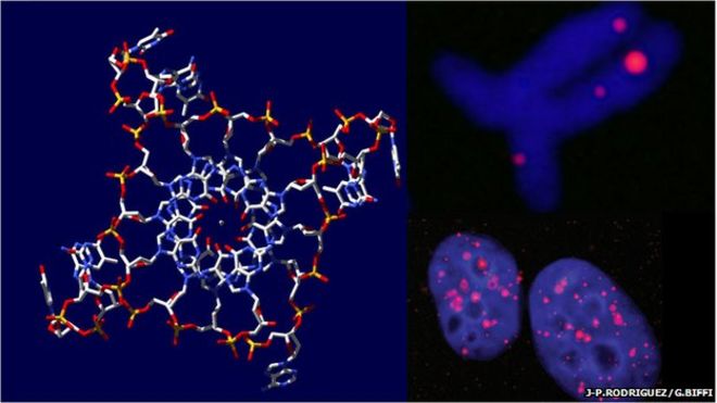
It’s been 67 years since Watson and Crick first established that DNA forms a double-stranded helix. Every now and then it is identified that the double-stranded DNA can double up, to form a special quadruple stranded helix form. 

Previously, quadruple DNA has been developed synthetically but was only seen in a curious point of view. Around 2013, scientists for the first time noticed quadruple DNA in human cancer cells. Today another milestone was set, when scientists for the first time have identified the peculiar DNA in healthy human cells. 

“We’ve undoubtedly demonstrated that the quadruple-strand DNA forms in living cells,” says Marco Di Antonio at Imperial College London. “This forces us to rethink the biology of DNA.”

How does quadruple DNA form?

The DNA molecule is made up of 4 nucleobases- guanine, adenine, cytosine, guanine, and thymine, which can configure themselves in multiple ways. 

At times, it happens so that the DNA can double up to form quadruple helix DNA, formed by sequences rich in guanine. Guanine is the only known nucleobase that is capable of forming bonds with each other. The quadruple DNA usually occurs helically in nature and contains the guanine bases stacked on top of each other.

The unimolecular regions are usually seen at the ends of the chromosome/ telomeric regions. This is usually seen in microbes, oncogenes and now in healthy human cells.

Time to rethink the biology of DNA:

The DNA was viewed by Di Antonio and team from the University of Cambridge. The team viewed the DNA within the human cell by attaching a novel fluorescent marker.

Quadruplexes, in the primary region of the DNA, act as an “on” and “off” switch particularly for the genes associated with cancer. Previous research suggests that the quadruple DNAs can be used as molecular targets to diagnose cancer at early stages. 

Although Di Antonio and the team are aware of the DNA function, it is still a mystery on how a cell knows where to express the gene or decrease its activity. The researchers hypothesize that the quadruple structure may hold the door open to facilitate the reading of the genetic code. 

“There is a sort of crosstalk between the formation of quadruplex DNA and epigenetic markers,” says Di Antonio. “The quadruplex form is an epigenetic mark in its own right.”

Concluding remarks:

This recent discovery adds to the evidence that the quadruple portions are a normal part of DNA regulation and function. It has also made us realize that we need to rethink our whole biology and that our view of double-stranded DNA may be out of date. 

Though the new discovery can be assumed to be a native state of DNA, it is another example proving that DNA is not a fixed shape or structure. 

Discovery of ‘on-off’ switch in plant immune defense

Living systems are known to be equipped with self defense mechanisms, and plants are no exception. For the first time, experts find a novel “on-off switch” in plant defense. 

The main purpose of an immune system is to protect the host from any harm. All living organisms are equipped with an immune system, and plants are no exception. Plants are home to rich sources of nutrients attracting many organisms including fungi, bacteria and vertebrates. Although plants lack an immune system as complex as humans, they do have stunning forms of chemical, structural and protein defense that experts are trying to understand. 

Plant immune defense:

Plants immune defenses are designed to detect any living organism and stop them from causing any extensive damage. Plants are equipped with multi layers of surveillance mechanisms that are capable of recognising potentially dangerous pathogens. 

The defense mechanisms are specifically programmed and operate through a complex network. They are timed for effective and rapid response against the predators, yet at the same time are controlled to avoid any damage to the host. 

The novel discovery:

Keini Dressano, Alisa Huffaker and team from the University of California San Diego, have recently discovered a novel ‘on-off’ switch in the plant defense system. 

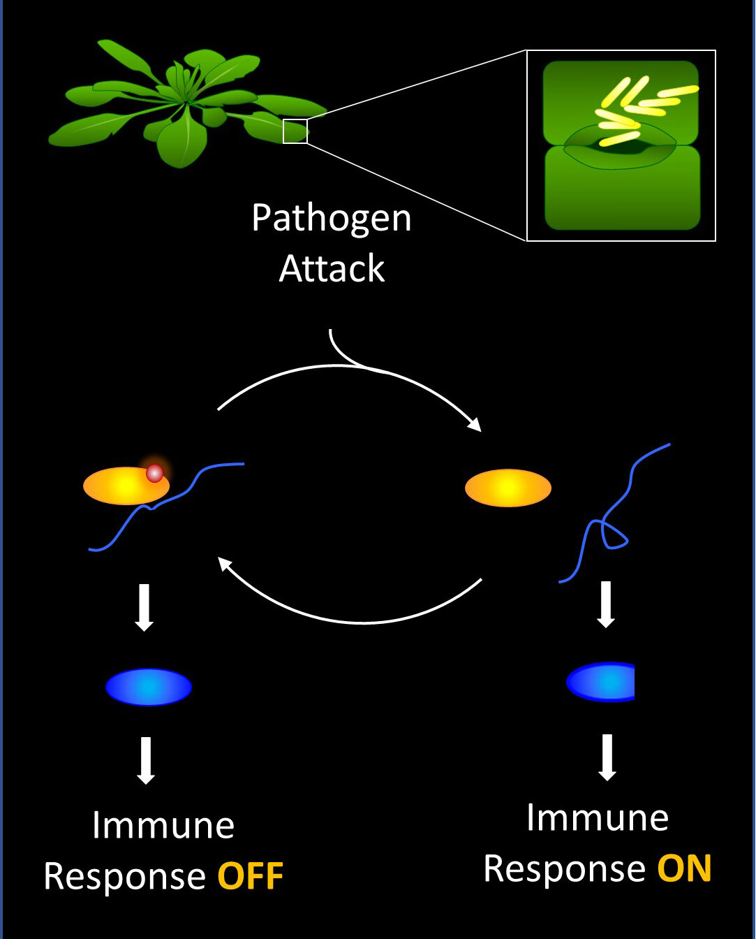
The new switch mechanism is an RNA binding protein that can help turning on immune responses a few minutes after being attacked. They also observed that it was capable of switching off the immune response hours later, to prevent self inflicting damage.

“These findings have provided new insights into how the complex intricacies of plant immune responses are orchestrated to successfully fight off pathogens, and lay a path forward for improving plant disease resistance to ensure future food stability,” said Huffaker, an assistant professor in the Section of Cell and Developmental Biology.

Science behind the discovery:

The novel discovery was found in arabidopsis plants, and it was found to control splicing of the mRNA transcripts that encode signalling protein regulators if the plants defense system.

According to the researchers, a simple chemical modification of the RNA binding protein was seen to reverse mRNA splicing that usually keeps the immune response deactivated. To turn on the immune response, a second chemical modification returns the mRNA splicing to normal resuming the inactivation process. 

“This work went beyond simply identifying a new regulator of plant immunity,” said Huffaker, of the detailed mechanisms uncovered. “We discovered specific chemical modifications that control regulatory function, transcriptional targets of the regulator, differential splicing of the targets and precise effects of splicing on both target function and overall plant immune responses and disease resistance.”

Source

Use of nanoparticles in drug carrier systems

Since ancient times, plant-based natural products have been commonly used as medicines. Nearly as much as 25% of major pharmaceutical compounds are derived from natural resources. They exhibit fascinating chemical diversity and biological properties with minimal toxicity.

Despite the advantages, companies are hesitant to invest more in natural delivery systems, due to concerns such as size, shape, and biocompatibility. The use of large materials in drug delivery includes many risks such as solubility and poor absorption. Such problems gave rise to nanotechnology, currently revolutionizing the field of medicine. 

Nanotechnology is an advanced field of nanoparticles being used in the science field at a molecular level. Nanoparticles are as small as 10nm to 1000nm, usually preferred to be less than 200nm. This helps them to move freely inside an organism when compared to other materials. Lately, the use of nanoparticles in drug delivery systems is speculating a lot of discussions.

Nanoparticles in drug delivery:

Currently, one of the challenges faced by researchers is to deliver a drug to an accurate site without causing any potential side effects to other healthy organs. This is especially important for cancer treatment, where the tumor is a distant metastasis in various organs. Nanoparticles employed, have specific properties to overcome the above limitation. These include their small size, ability to penetrate cell membranes, binding and stabilizing proteins. Nanoparticle entrapment of drugs is performed to make sure the drug is delivered to the target tissue supported with a controlled release. 

The idea of using nanoparticles with natural compounds is very attractive in recent times. Natural compounds have a lot of properties that are enhanced by combining with nanoparticles. These properties include targeting a specific tissue and controlled release of the drug. 

Metallic, inorganic, and polymeric nanoparticles are some of the frequently used types in performing a target-specific drug delivery system. In particular, drugs with poor solubility need the aid of these nanoparticles. 

The efficacy of the use of nanoparticles varies according to the material, size, and other biodegradability properties. Due to their biocompatibility properties, various synthetic polymers such as polyvinyl alcohol and poly-L-lactic acid, and natural polymers such as chitosan and alginate are broadly used in fabricating nanoparticles. 

Polymeric nanoparticles can be divided into nanocapsules and nanospheres, both of which are proven to be attractive drug delivery systems. The use of an optimal nano-drug delivery system is based upon the properties of the drugs chosen for the treatment. 

Possible hazards:

When combining nanotechnology in medicine, toxicity exhibited by nanoparticles cannot be ignored. For pharmaceuticals, specific drug formulations can be put in place for clinical efficacy and minimizing toxicity. 

Nanoparticles are known for their unique surface properties, and since it is the contact layer with the body tissue, it should be evaluated from a toxicology standpoint. Although a lot of tests and procedures are available to evaluate the material, it cannot be assumed that these tests will be precise in detecting all risks. This will most likely depend on the origin of the materials- biological or non-biological.

Recently, researchers are exploring the ideas of combining nanoparticles and natural products to minimize toxicity. Both individually have already been in use for several years, but have their own limitations. The greenway of formulating nanoparticles is widely encouraged as it lowers the hazardous constituents during the synthesis process. By using green nanoparticles, the chances of side effects can be minimized. Furthermore, by changing the shape, size, and hydrophobicity we can further enhance the bioactivity of nanoparticles.

Future of nanoparticles in the medicinal field:

Nanoparticles in the medicinal field is currently one of the fascinating areas of research out there. In the future, by the looks of it, the field of cancer looks to benefit the most from nanoparticles. By using various types of nanoparticles, it is possible to transport an accurate amount of drug to the tumor cells without causing harm to other cells. The application of nanoparticles in medicine will definitely be the future trend in diagnosis and treatment research. 

More research needs to be performed to achieve a more consistent drug loading and release capacity. , progress is being made in the use of metal-based nanoparticles like gold and silver in diagnosis and therapy areas of research. 

Nanotechnology is truly a multidisciplinary science beneficial to drug delivery systems. Despite the overwhelming advantages, its actual impact on the healthcare system is quite limited. In the future, a more conceptual understanding of biological response to nanoparticles must also be studied. Ultimately researchers should be able to deliver drugs for a long period of time with great precision and controlled release.

What does it mean when an article is peer-reviewed?

What does it mean when an article is peer-reviewed?

A top-quality research paper is always a combination of original research and good writing. Whether a student or a renowned research scientist, writing and submitting a research paper is not an easy task. It goes through several rounds of editing and review, both time-consuming and exhausting.

What is a peer review?

Reviewers are known to play an important role in any scholarly publishing. Peer review is a long, arduous process of subjecting a scholarly or research work to the scrutiny of others who are fellow experts in the field. The concept of the peer review was conducted long before the first known scholarly journal. For centuries the process has been used as a method to evaluate written work. 

Peer review is primarily conducted for two reasons. Firstly, it acts as a filtered process to ensure only high standard research and content are published by validating the originality and significance of the study. Secondly, it is intended to encourage the authors to meet the high standards and to ensure any unwarranted claims or personal views are not published.

An overview of the peer-review process:

The process usually begins right after the author has completed the research and has finished the manuscript. A manuscript usually contains the purpose of the study, design, results, and conclusions. The paper is then submitted to a suitable journal of choice usually relevant to the field of study. The paper is then reviewed by the journal editors to ensure the subject matter is in the same line as the journal. 

As the next step, the paper is sent to certain experts in the field related to the study for a peer-review process. Peer reviewers are also popularly known as referees. Editors work includes that the peer review process takes place fairly and in a timely and effective manner. Additionally, they must also monitor if there are any conflicts of interest involved during the review process. 

As a first step, the reviewer first reads the whole paper thoroughly to carefully scrutinize the validity of the science, the quality of the study, and the methods used. Additionally, the findings from the study are evaluated and its contribution to the field advancement.

Furthermore, the reviewers also identify any scientific flaws or if any references are missing or inappropriate. They later give recommendations to the editor whether the paper should be accepted or not. If the paper is good enough with minute errors, the editor acts as a mediator between the referee and the author to make the required corrections if any. 

If accepted, as per the referees’ recommendation the after goes to the production stage, where it is properly formatted by the editors according to the journal standards.

Types of peer-review:

The peer-review process takes many forms. It must, therefore, be checked which type is employed by a journal before working on a paper. 

Single-blind review:

In this type of review, the reviewers/referees’ name is usually hidden to prevent any impartial decisions or influence by the authors. It is a traditional method of reviewing and is most popularly used. Sometimes, reviewers may misuse their anonymity and portray unnecessary criticisms when reviewing work. 

Double-blind review: 

In this process, both the author and the reviewer are hidden. This way there is no bias like the author’s sexuality, academic status, or country of origin. This type of review is conducted to make sure that articles written by renowned scientists or authors are considered on the basis of their content research rather than their reputation. 

Triple blind review:

In this peculiar case, all three people involved: editor, reviewer, and author are unknown to each other. The paper is anonymized even before the submission stage and is handled this way to keep away any bias towards the author. 

Peer review has become an essential tool in assisting the journal editor in selecting high quality, credible, novel, and highly interesting scientific papers. Though not all peer-reviewed processes are smooth or accurate, a more suitable and tedious process hasn’t been discussed or developed. 

A more transparent review:

As the name suggests, this type of review process has full transparency. Both the authors and the reviewer’s names are open. It is popularly believed that this is the best way to prevent any plagiarism, malicious comments, and to encourage honest, open reviewing. A few of them feel it is a less honest process, which may cause the reviewer to hold back his comments due to fear or politeness. 

Peer review has become an essential tool in assisting the journal editor in selecting high quality, credible, novel, and highly interesting scientific papers. Though not all peer-reviewed processes are smooth or accurate, a more suitable and tedious process hasn’t been discussed or developed. 

It is high time journals start looking at options to make this process automated. This ensures a foolproof system ensuring to release high quality and error-free papers into the scientific community. 

MYC Drives Temporal Evolution of Small Cell Lung Cancer subtypes by reprogramming Neuroendocrine Fate

MYC Drives Temporal Evolution of Small Cell Lung Cancer subtypes by reprogramming Neuroendocrine Fate

Small Cell Lung Cancer (SSLC) is a very particular lethal malignant cancer type for which effective therapies are urgently needed. Small cell carcinomas are usually centrally located, arising from the bronchus, with a small number of peripheral lesions. They obstruct most of the airways through circumferential compression. 

Around 15% of lung cancers are classified under SCLC. Recent studies indicate that SCLC can be split into 4 major subtypes, based on the expression YAP1, ASCL1, NEUROD1, or POU2F3. Treatments for these subtypes are not yet standardized, due to the lack of information on their physiology. 

Now scientists have summarised their new findings “MYC Drives Temporal Evolution of Small Cell Lung Cancer subtypes by reprogramming Neuroendocrine Fate” in Cancer Cell about the physiology of SCLC subtypes, which could potentially pave the way to study and treat the disease. 

Historically SCLC has been treated as a single disease. The disease exhibits genetic loss of tumor suppressors along with the expression of MYC, MYCL, or MYCN. Large scale gene expression analyses suggest that SCLC subtypes have distinct vulnerabilities which need to be understood to improve treatment. Subtype SCLC-ASCL1 which comprises 70% of the tumors is a regulator of neuroendocrine (NE) fate. Using genetically engineered mouse models (GEMM) it was found that ASCL1 is important for tumor development and that MYCL was highly expressed in this subtype. Contrastingly the other SCLC subtypes constituting 30% found to overexpress MYC and exciting low non-NE cell fate. Research shows that Myc expression drives a non-NE SCLC in GEMMs, however, the relationship between the subtypes SCLC-A and ACLC-N and involvement of MYC in driving other subtypes is still unknown. 

In this paper, the author and his team investigate MYC origins and its relationship with the SCLC subtypes. Functional data from the study suggest that MYC is behind the evolution of SCLC subtypes. In context to the times’ suppressor loss, MYC promotes temporal evolution from SCLC-A to SCLC-N to SCLC-Y in-vivo. The results from the study also revealed that MYC does not work alone and requires the help of the NOTCH signaling pathway to drive tumor progression. 

 The study further revealed that both MYCL and MYC are not functionally redundant in SCLC. They correlate with distinct gene expression and localize to super-enhancers. MYC and MYCL have the ability to change a cell morphology fate, molecular subtype, and influence drug sensitivity. 

Different SCLC subtypes inhibit different targeting drugs and considering this, dynamically evolving tumors are termed as moving therapeutic targets. Over the years many targeted therapies have failed to treat SCLC like chemotherapy. The authors of this study speculate that chemotherapy has remained the most effective due to its non-specificity among subtypes that evolve. The findings from these studies demonstrate that SCLC and other similar cancer types would benefit from a combination or customized therapies.

Source