Adapting to survive: How Candida overcomes host-imposed constraints during human colonization

Adapting to survive: How Candida overcomes host-imposed constraints during human colonization

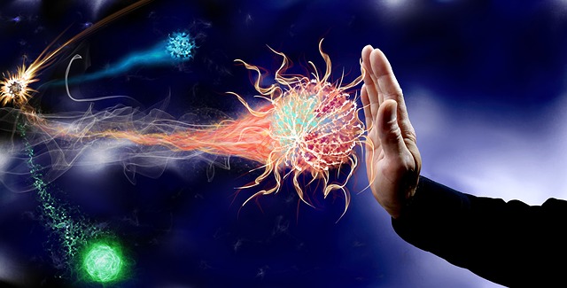
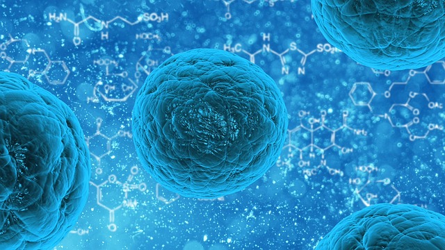
The human body is well known to host a large number of microbes, mostly harmless but when triggered might turn virulent. A large fungal ecosystem resides inside a human body mainly including Candida species, constituting a large part of the human body’s microbial flora. Usually asymptomatic, Candida forms small colonies, but when triggered such as environmental change, can potentially help the microbes to break barriers and cause life-threatening diseases.

Though multiple numbers of antifungal drugs are available, it is recently found that Candida species is capable of building resistance against the drugs by forming biofilms. The article further talks about the environment within the host body paving way to such resistance. 

Within the human host, Candida is capable of changing morphology and functions according to the change in the environment it resides in. Several factors play a role including temperature, ph, and oxygen supply. Candida depending on the environment can take forms such as hyphae, budding, or even pseudohyphae. 

Another crucial role played in a microbial existence within the human host is nutrients availability. It is reported that microbes thrive in the area of high glucose content. When deprived of glucose is when microbes turn to another source of nonfermentable nutrients. Research performed in labs using Candida flora has reported that in the presence of glucose the microbe is known to morph into hyphae and promote antifungal resistance. 

The limitations of micronutrients such as iron magnesium, and copper are known to limit the growth of invading microbes. But this is quite tricky as micronutrients are needed both by the host and microbes in functioning such as biochemical and cellular functions. 

It is very well known that oxygen and ph levels vary within every niche in the human body. While some are alkaline and high on oxygen concentration others are hypoxic and acidic. Candida microbes being versatile they are, can adapt their cell walls according to the change in ph. It is also interesting to note that Candida microbes thrive under hypoxic conditions, inducing their hyphal growth and causing immune evasion. 

The above has described the flexibility of the microbes to overcome multiple constraints faced in the host body. This ability of Candida helps it to form colonies and invade niches around the body. Another strategy imparted by the microbes is biofilm formation against the host body or biomedical devices. Biofilms consist of a 3D community of adherent cells with different biological properties. These cells are embedded in the ECM, which helps in maintaining the overall integrity of the biofilm. The ECM also acts as a protective barrier against any drug invasion. These features play a crucial role in Candida microbes resistance against antifungals and biomedical devices. 

With the emergence of resistant Candida species, the need to develop new antifungals is inevitable. Research using an in vivo model to mimic the host conditions is giving close insights to unravel the mysteries of the microbes. These approaches are paving the way to novel therapeutic vaccines and anti-fungal treatments, enhancing the body’s ability to fight off the infections. 

Source

Artificial intelligence-enabled rapid diagnosis of patients with COVID-19

Artificial intelligence-enabled rapid diagnosis of patients with COVID-19

Since December 2019, multiple cases of pneumonia due to unknown reasons have emerged in Wuhan, China. Through testing multiple patient samples, scientists extrapolated a new coronavirus termed COVID-19. With no FDA approved therapeutics or treatment available for the disease, diagnosis plays an important role in containing COCVID-19, giving a path to the rapid implementation of control measures to limit the spread. With the disease spreading to almost 100 countries, a million cases have been confirmed worldwide to date. Imaging is one of the main principles used in diagnosing and evaluating the disease, with the final diagnosis depending on reverse transcriptase-polymerase chain reaction (RT-PCR). 

In response to the growing number of COVID-19 cases, there is currently a shortage of diagnostic kits worldwide. Multiple industries are coming forward to develop rapid, easy to use diagnostic kits to facilitate testing. However before these kits can be commercialized, they must be tested and validated. With the current available tests taking almost 2 days to complete and produce a result, serial testing is required to rule out any negative cases. Additionally, it is a mystery as to whether an RT-PCR is a gold standard and whether a false positive/ negative result is common. The above reasons highlight the need for alternative testing methods to produce rapid and accurate results to identify, isolate, and treat the affected people. 

Chest computed tomography is also a much-used valuable component in testing COVID-19. With some of the patients showing early-stage symptoms in radiological finding, limits the CT ability to differentiate between a positive and negative case. In this current study, the authors have used Artificial Intelligence (AI) algorithms to help in integrating CT scanning in finding the symptoms of the virus, exposure history and reliable lab testing to rapidly diagnose the patients affected with COVID-19. 

A trial was performed on 905 patients diagnosed using RT-PCR and next-generation RT-PCR and around 46% (419) people were declared positive for COVID-19. Parallelly in a test set of 279 participants, the AI system managed to achieve accuracy to about 92% of the population and had equal or even better sensitivity than a senior radiologist. The AI system also improved the detection of COVID-19 positive patients with negative CT scans, identifying 17 out of 25 participants who were tested positive via RT-PCR but negative with normal CT scans. In comparison, the radiologists’ declared the said 17 participants to be COVID negative. 

AI shows signs of analyzing huge amounts of data quickly, a quality that is much needed in the current pandemic. A major limitation of the above study is the small sample size, with available CT scans and clinical history data, the AI system can help in diagnosing COVID-19 patients rapidly. Though a promising tool, further data collection is required to test the generalization of AI mapping on other patient populations.

Source

Scientists Help Immune System Find Hidden Cancer Cells

Scientists Help Immune System Find Hidden Cancer Cells

Cancer is a widely studied topic in bioscience research but yet remains to be mostly unknown and difficult to treat. However, the recent developments in cancer research over the past decade have helped the scientific community to come up with effective treatment methods. But still, one of the most common problems faced in treating cancer cells is difficulty in locating them for efficient targeting. Recently, scientists from Yale University have developed a new system that can help our immune system find the hidden cancer cells and kill them. The research has been published in Journal Nature Immunology.

Why this study holds importance?

It is a known fact that there exists a number of immunotherapies for treating cancers. But these therapies have certain shortcomings as they either don’t work on all patients or are inefficient in different cancer types. The major reason behind this is the failure of these therapies in identifying the cancer cells which reduces their effectiveness. This highlights an urgent need for a more targeted approach that can help curb the menace of cancer.

The development of a new system by scientists in the present study is considered to overcome the drawbacks of the earlier immune therapies. Researchers report that upon testing the new system in mice it has shown positive response against the melanoma, triple-negative breast, and pancreatic tumors, even for those tumors which are situated at a distant location from the primary tumor.

“This is an entirely new form of immunotherapy,” said Sidi Chen, senior author.

How the new system – MAEGI works?

The researchers developed a new system to target the cancer cells which combines the viral gene therapy and CRISPR based gene-editing technology. Unlike the traditional method of searching and making edits at the DNA level by incorporation of new genes, the present system uses a much more targeted approach.

The new system named as MAEGI stands for Multiplexed Activation of Endogenous Gene as Immunotherapy. This system works by searching numerous cancer-causing genes, marking their location by mimicking GPS and subsequently intensifying the signal of these locations for precise targeting.

For instance, you can consider that the new system dresses up the tumor cells in a unique manner that can be easily identified by the immune system of our body and eventually eliminate them. For this, the cold tumors cells lacking any immune cells are converted into hot tumors cells which are packed with tons of immune cells.

“And once those cells are identified, the immune system immediately recognizes them if they show up in the future,” Chen said. The new system, in theory, should be effective against many cancer types, including those currently resistant to immunotherapy, he said.

The researchers will be further optimizing this system to make the manufacturing process easier. Once optimization is done it will be subjected to clinical trials in potential cancer patients.

No wonder that cancer is a rising menace in the present world. Though many therapies are available we still need highly effective methods to treat cancer. The development of the immunotherapy-based system has given rise to a ray of hope for a more effective and proficient treatment that not only treats primary tumor cells but also the distant ones. Let’s look forward to this new development and hope for the best outcomes in subsequent clinical trials.

Source 1

Source 2

An in situ-Synthesized Gene Chip for the Detection of Food-Borne Pathogens on Fresh-Cut Cantaloupe and Lettuce

Food-borne pathogens are one of the major reasons behind endangering the life and safety of people across the globe. Fresh foods are specifically more vulnerable to these pathogens, making it crucial to have a very efficient food safety surveillance technology. The development of such technology will help in offering rapid detection of food-borne pathogens. In the present study, researchers developed an In-situ synthesized gene chip for the detection of the food-borne pathogen. Here the researchers first identified and screened the target genes by comparing the sequences of common food-borne pathogens like Salmonella, Vibrio parahemolyticus, Staphylococcus Aureus, Listeria monocytogenes and E.coli 0157:H7 from the NCBI database. Unique tilling array probes were designed that helps to target the selected genes in an optimized hybridization system. The resultant assay showed high specificity along with strong amplification signals. The results were highly accurate with a detection limit of approximately 3 log cfu/g without culturing. The detection time for the five target food-borne pathogens on the fresh-cut cantaloupe and lettuces was found to be 24 hours. This highlights the great efficacy of the detection system to rapidly monitor the pathogens on the fresh food items. Such a system can be easily incorporated as an efficient food surveillance system for checking the logistical distribution chain, the food at the processing stage, cleaning condition at the food manufacturing plants, transport, sales and more. The technology is considered valuable as it supports the safety of fresh agricultural products, reducing the overall wastage of food due to infectious pathogens.