Groundbreaking research by Monash University paves the way to a possibility of better monitoring and treatment of the cystic fibrosis lung disease
For years, cystic fibrosis (CF) has known to be a serious hereditary condition that causes severe damage to the respiratory and digestive systems. The damage starts with a build-up of thick mucosal kind of fluid in the organs especially lungs.
In particular cystic fibrosis affects the cells that produce digestive enzymes, sweat, and mucus. Usually, these secretions are thin and watery, which function as lubrication for various organs and tissues. However, when affected with CF, due to faulty genes the person experiences thick fluid build-up, clogging up the passages and ducts.
Early diagnosis and treatment are important for improving the quality and the length of the affected life. Currently available lung assessment tools have many drawbacks, especially the inability to accurately identify the origin of the changes seen in lung health.
Monash University research team announced the results of the World’s first research promising a possibility of better diagnosis and treatment of cystic fibrosis using X-ray velocimetry.
What is X-ray velocimetry?
A phase-contrast X-ray imaging makes use of the refractive properties of materials to produce high definition soft tissue images. Recently X-ray velocimetry (XV) has caught the eye of researchers. In particular, XV is used to study the airflow through the lungs and the technique is based on particle image velocimetry. Particle image velocimetry (PIV) is a well-established technique known for years.
XV is known to provide high quality, non-invasive, real-time images of the airflow through the lungs. The X-ray was first designed and developed by 4DMedical, in hope for clinical use. The technology has also recently been approved by the FDA for its all respiratory indications in adults.
All about the study:
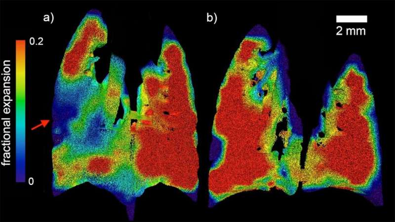
An effective assessment of the lung cystic fibrosis should be capable of capturing its patchy nature and fluid buildup. This is in particular important for detecting the disease at early stages.
With the help of XV, a multidisciplinary collaboration of engineers, physicists, and clinicians were able to measure real-time airflow through the lungs.
The research was led by one of the University’s leading scientists Dr. Freda Werdiger. The study revealed that by using XV, it was possible to pinpoint the precise locations where there was an obstruction of airflow in the lung of a cystic fibrosis patient.
“In this study, we present two developments in XV analysis. Firstly, we show the ability of laboratory-based XV to detect the patchy nature of CF-like disease in affected mice. Secondly, we present a technique for numerical quantification of that disease, which can delineate between two major modes of disease symptoms,” Dr. Werdiger said.
This particular model provides a very simple, easy top interpret approach, which in the future can be readily applied to large quantities of data generated in XV imaging.
What does the future hold?
The success of XV lies in its capability of drawing reliable and quantitative measures, and the above study shows how that can be accomplished.
The researchers of the study recommend that this promising technique should be applied to the numerical characterization of CF lung disease. The analysis can be applied in a straight forward fashion with minimal manual labour required.